Investig Magn Reson Imaging.
2019 Jun;23(2):172-174. 10.13104/imri.2019.23.2.172.
Recurrent Painful Ophthalmoplegic Neuropathy: a Case Report
- Affiliations
-
- 1Department of Radiology, Jeju National University Hospital, Jeju National University School of Medicine, Jeju, Korea. hoklee33@gmail.com
- 2Department of Neurology, Jeju National University Hospital, Jeju National University School of Medicine, Jeju, Korea.
- KMID: 2452533
- DOI: http://doi.org/10.13104/imri.2019.23.2.172
Abstract
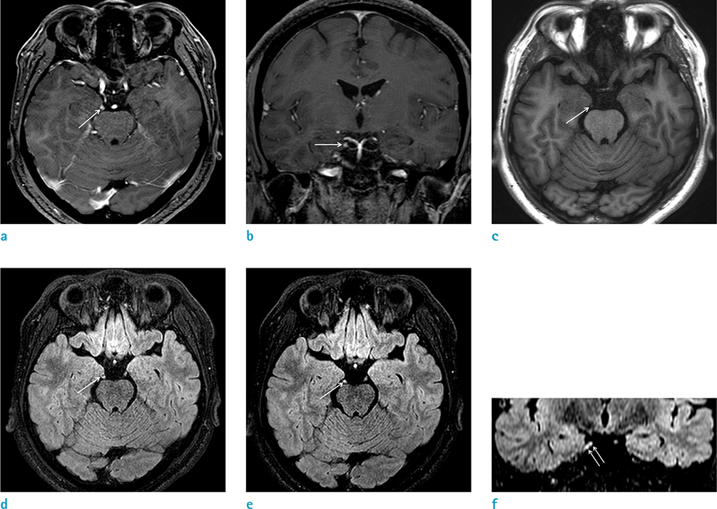
- Upon review, it is noted that recurrent painful ophthalmoplegic neuropathy (RPON) is a rare neurological syndrome characterized by recurrent unilateral headaches and painful ophthalmoplegia of the ipsilateral oculomotor nerve. As seen on brain MRI, thickening and enhancement of the oculomotor cranial nerve can be observed in these cases. We experienced a case of RPON in an adult patient who showed thickening and enhancement of the oculomotor nerve on gadolinium-enhanced 3D-FLAIR image. The authors report a case of RPON with a review of the literature.
MeSH Terms
Figure
Reference
-
1. Gelfand AA, Gelfand JM, Prabakhar P, Goadsby PJ. Ophthalmoplegic "migraine" or recurrent ophthalmoplegic cranial neuropathy: new cases and a systematic review. J Child Neurol. 2012; 27:759–766.2. The international classification of headache disorders 3rd edition. International headache society web site. 2018. Accessed June 12, 2019. https://www.ichd-3.org/13-painful-cranial-neuropathies-and-other-facial-pains/13-10-burning-mouth-syndrome-bms/.3. Bharucha DX, Campbell TB, Valencia I, Hardison HH, Kothare SV. MRI findings in pediatric ophthalmoplegic migraine: a case report and literature review. Pediatr Neurol. 2007; 37:59–63.
Article4. Mark AS, Casselman J, Brown D, et al. Ophthalmoplegic migraine: reversible enhancement and thickening of the cisternal segment of the oculomotor nerve on contrast-enhanced MR images. AJNR Am J Neuroradiol. 1998; 19:1887–1891.5. Kivrak AS, Nayman A, Paksoy Y, Ozturk S. Magnetic resonance findings in recurrent painful ophthalmoplegic neuropathy with reversible enhancement of oculomotor nerve. Eur J Gen Med. 2015; 15:64–66.6. Miglio L, Feraco P, Tani G, Ambrosetto P. Computed tomography and magnetic resonance imaging findings in ophthalmoplegic migraine. Pediatr Neurol. 2010; 42:434–436.
Article7. Park JW, Moon HS, Kim JM, Lee KS, Chu MK. Chronic daily headache in Korea: prevalence, clinical characteristics, medical consultation and management. J Clin Neurol. 2014; 10:236–243.
Article8. Lim HK, Lee JH, Hyun D, et al. MR diagnosis of facial neuritis: diagnostic performance of contrast-enhanced 3D-FLAIR technique compared with contrast-enhanced 3D-T1-fast-field echo with fat suppression. AJNR Am J Neuroradiol. 2012; 33:779–783.
Article9. Naganawa S, Sugiura M, Kawamura M, Fukatsu H, Nakashima T, Maruyama K. Prompt contrast enhancement of cerebrospinal fluid space in the fundus of the internal auditory canal: observations in patients with meningeal diseases on 3D-FLAIR images at 3 Tesla. Magn Reson Med Sci. 2006; 5:151–155.
Article
- Full Text Links
- Actions
-
Cited
- CITED
-
- Close
- Share
- Similar articles
-
- A Case of Ophthalmoplegic Migraine Developed in Infancy
- Ophthalmoplegic Migraine: A Case Report
- An Adolescent Case of Recurrent Episodes of Ophthalmoplegic Migraine
- Recurrent Painful Ophthalmoplegic Neuropathy in an 8-Year-Old Boy
- A Case of Autonomic Dysfunction and Painful Sensory Neuropathy in Sjogren's Syndrome


