Pelvic Actinomycosis Mimicking Malignancy of the Uterus: a Case Report
- Affiliations
-
- 1Department of Radiology, Soonchunhyang University College of Medicine, Seoul Hospital, Seoul, Korea. jy0707hwang@schmc.ac.kr
- 2Department of Pathology, Soonchunhyang University College of Medicine, Seoul Hospital, Seoul, Korea.
- KMID: 2452527
- DOI: http://doi.org/10.13104/imri.2019.23.2.136
Abstract
- Pelvic actinomycosis is an uncommon infectious disease. It induces a chronic, suppurative illness characterized by an infiltrative and granulomatous response and, thus, the clinical and radiologic findings may mimic other inflammatory and neoplastic conditions. A 56-year-old female with a long-standing intrauterine device was diagnosed with pelvic actinomycosis manifesting as a large uterine mass with locally infiltrative spread into surrounding tissue that mimicked uterine malignancy. Actinomyces israelii infection was confirmed with a surgical specimen, and the patient was treated with antibiotic medication. Pelvic actinomycosis must be included in the differential diagnoses of patients with an infiltrative pelvic mass extending across tissue planes or in patients with findings of multiple microabscesses, particularly in a patient with an intrauterine device, even the lesion primarily involves the uterus.
Keyword
MeSH Terms
Figure
-
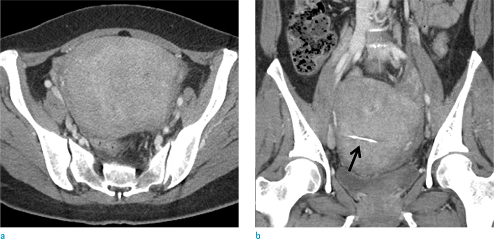
Fig. 1 A contrast-enhanced abdominal axial (a) and coronal (b) reformatted CT images on initial presentation showed diffuse enlargement of the uterus with a poorly defined, heterogeneously enhancing lesion. There was an IUD in situ (arrow).

Fig. 2 A follow-up contrast-enhanced abdominal axial (a) and coronal (b) reformatted CT images 3 months after initial presentation showed enlarged heterogeneously enhancing uterine lesion with multiple focal areas of low-attenuation. There was an IUD in situ (arrow). The adjacent sigmoid colon showed mild luminal narrowing with wall thickening, and there was soft tissue stranding in the surrounding mesocolon (arrowhead).
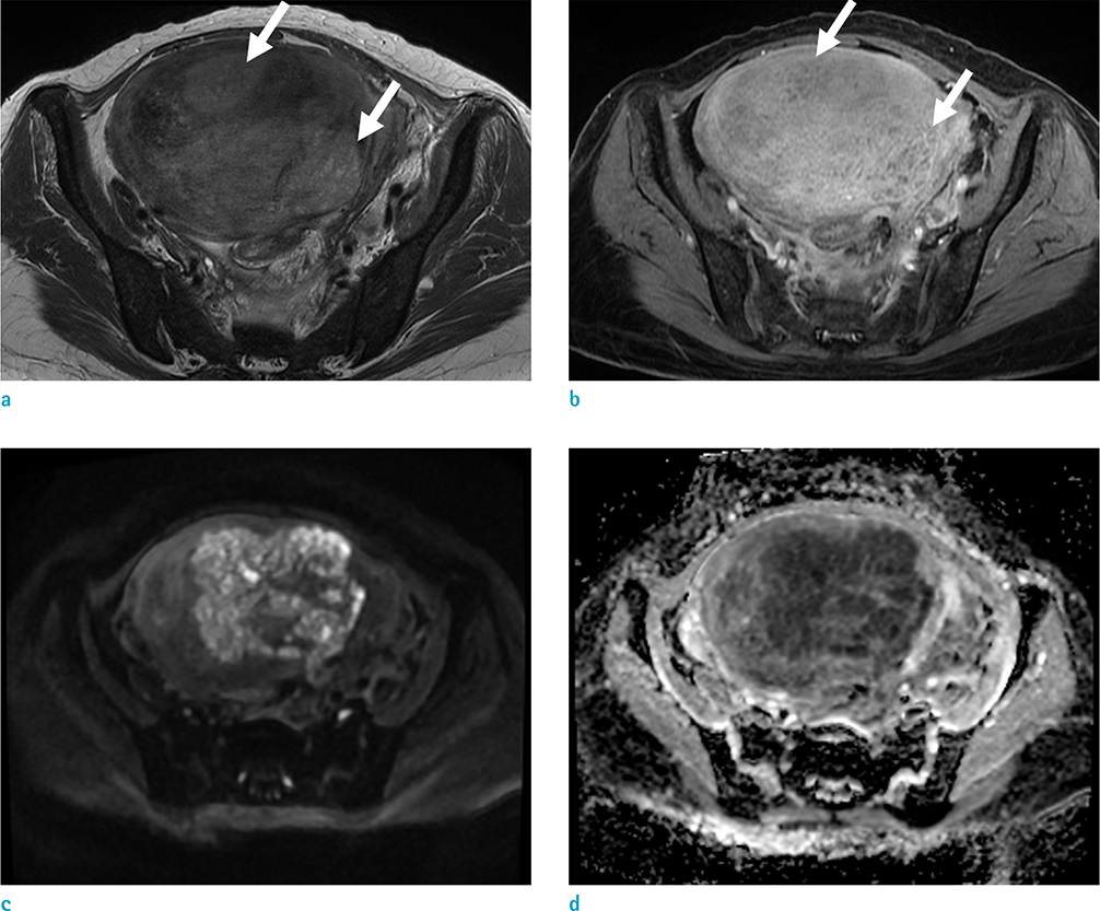
Fig. 3 (a) MRI revealed mild high signal intensity uterine mass with a poorly defined margin, small cystic foci on the axial T2-weighted image (arrows), and (b) heterogeneous enhancement with multiple, tiny, hypointense foci with ring-like enhancement on gadolinium-enhanced fat-suppressed T1-weighted image (arrows). The lesion showed diffusion restriction on diffusion weighted imaging (c, d).
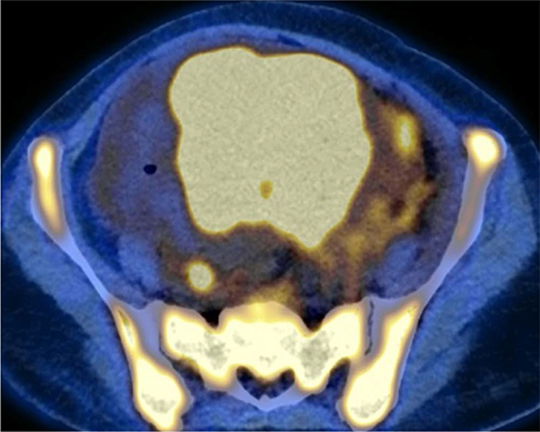
Fig. 4 On 18F FDG PET/CT, the lesion was highly avid (SUVmax = 20.0 g/ml). Diffuse hypermetabolism was also seen in the bone marrow cavity of the whole skeletal system. FDG PET/CT = fluoro-deoxyglucose positron emission tomography/computed tomography, SUVmax = maximum standardized uptake value.
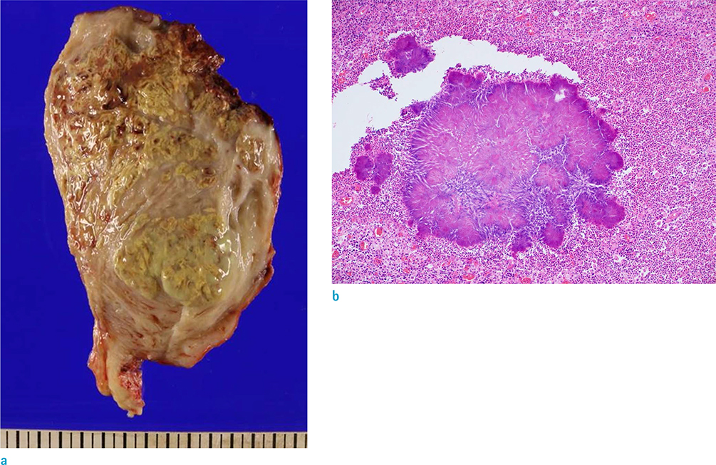
Fig. 5 On cut sections of the uterine gross specimen (a), a huge globoid uterine corpus revealed a markedly thickened myometrium showing multifocal, ill-defined abscesses and pus-like material. (b) Histopathologic features showed a sulfur granule in an inflammatory exudate (Hematoxylin & Eosin stain, × 100).
Reference
-
1. Montori G, Allegri A, Merigo G, et al. Intra-abdominal actinomycosis, the great mime: case report and literature review. Emerg Med Heal Care. 2015; 3:2.
Article2. Wagenlehner FM, Mohren B, Naber KG, Mannl HF. Abdominal actinomycosis. Clin Microbiol Infect. 2003; 9:881–885.
Article3. Ha HK, Lee HJ, Kim H, et al. Abdominal actinomycosis: CT findings in 10 patients. AJR Am J Roentgenol. 1993; 161:791–794.
Article4. Fiorino AS. Intrauterine contraceptive device-associated actinomycotic abscess and Actinomyces detection on cervical smear. Obstet Gynecol. 1996; 87:142–149.
Article5. Lely RJ, van Es HW. Case 85: pelvic actinomycosis in association with an intrauterine device. Radiology. 2005; 236:492–494.
Article6. Edwards DF, Nyland TG, Weigel JP. Thoracic, abdominal, and vertebral actinomycosis. Diagnosis and long-term therapy in three dogs. J Vet Intern Med. 1988; 2:184–191.7. Von Lichtenberg F. Infectious disease. In : Contran RS, Kumar V, Robbins SL, editors. Robbins pathologic basis of disease. 4th ed. Philadelphia: Saunders;1989. p. 383–384.8. Morland D, Hassler S. Case 219: Pelvic actinomycosis mimicking malignant tumor. Radiology. 2015; 276:304–308.
Article9. Alfuhaid T, Reinhold C. Pelvic actinomycosis associated with intrauterine device use: case report. Can Assoc Radiol J. 2003; 54:160–162.
- Full Text Links
-
- Actions
-
Cited
- CITED
-
- Close
- Share
-
- Similar articles
-
- A case of pelvic actinomycosis mistaken for advanced ovarian cancer
- A Case Of Pelvic Actinomycosis Complicated By Tuboovarian Abscess
- A case of pelvic actinomyosis presenting as an advanced ovarian malignancy
- Ureteral Obstruction Caused by Pelvic Actinomycosis
- Pelvic actinomycosis with hydronephrosis and colon stricture simulating an advanced ovarian cancer
