Vascular foramina of navicular bone: a morphometric study
- Affiliations
-
- 1Department of Anatomy, Mahatma Gandhi Medical College & Research Institute, SBV University, Puducherry, India. prathapamchandravani@gmail.com
- KMID: 2451233
- DOI: http://doi.org/10.5115/acb.2017.50.2.93
Abstract
- The navicular bone is supplied by more than one artery. The knowledge about the vascular foramina is important to understand the pathogenesis and management of navicular fractures. The objective of the present study is to analyze the morphology and morphometry of vascular foramina of dried human navicular bone in Indian population. The study was carried out by using 100 navicular bones (50 right and 50 left) collected from our institute and other medical institutes in and around Puducherry. The bones were macroscopically studied for vascular foramina with respect to its location, number, size, and shape. The data collected were statistically analyzed. The vascular foramina were present on dorsal, plantar, medial, and lateral surfaces of navicular bone. Kruskal-Wallis test followed by series of Mann-Whitney test for post hoc analysis showed the number of nutrient foramina observed on dorsal surface were significantly greater than those observed on the plantar (U=2,755, P=0.001), medial (U=43, P=0.001), and lateral (U=626.5, P=0.001) surfaces of the navicle. About 97.6% of foramina were circular and 2.5% were oval in appearance. About 96.7% of vascular foramina were <1 mm in size and 3.3% were ≥1 mm in size. Spearman's rank correlation coefficient done showed a strong, positive correlation between vascular foramina of <1 mm size and circular shape, which was statistically significant (r(s)=0.981, P=0.001). We believe the present study has provided additional information on the vascular foramina of navicular bone and useful to surgeons in foot surgeries.
Keyword
Figure
-
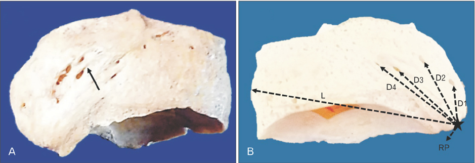
Fig. 1 Representative photographs showing the navicular bone. (A) Right navicular bone showing well-marked groove leading to vascular foramen (arrow) on dorsal surface. (B) Left navicular bone showing the measurements on dorsal surface of the navicular bone. RP, midpoint of navicular tuberosity; D1, the distance from the RP to the first vascular foramina; D2, the distance from the RP to the second vascular foramina; D3, the distance from the RP to the third vascular foramina; D4, the distance from the RP to the fourth vascular foramina; L, length of the navicular bone.
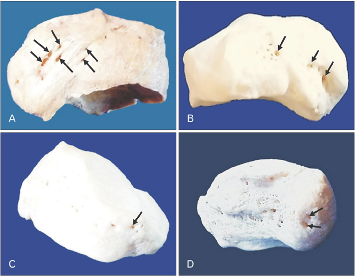
Fig. 2 Representative photographs showing different surfaces of navicular bone. (A) Right navicular bone showing the oval type vascular foramina (arrows) on dorsal surface. (B) Left navicular bone showing the circular type vascular foramina (arrows) on the plantar surface. (C) Right navicular bone showing the circular type vascular foramina (arrow) on lateral surface. (D) Left navicular bone showing the oval type vascular foramina (arrows) on medial surface.
Reference
-
1. Moore KL, Dalley AF, Anne MR. Clinically oriented anatomy. 6th ed. Philadelphia: Lippincott Williams & Wilkins;2010. p. 324.2. Mahadevan V. Ankle and foot. In : Stranding S, editor. Gray's Anatomy: The Anatomical Basis of Clinical Practice. 40th ed. Edinburgh: Elsevier Churchill Livingstone;2010. p. 1429–1462.3. Golano P, Fariñas O, Sáenz I. The anatomy of the navicular and periarticular structures. Foot Ankle Clin. 2004; 9:1–23.4. Eichenholtz SN, Levine DB. Fractures of the tarsal navicular bone. Clin Orthop Relat Res. 1964; 34:142–157.5. Saxena A, Fullem B, Hannaford D. Results of treatment of 22 navicular stress fractures and a new proposed radiographic classification system. J Foot Ankle Surg. 2000; 39:96–103.6. Torg JS, Pavlov H, Cooley LH, Bryant MH, Arnoczky SP, Bergfeld J, Hunter LY. Stress fractures of the tarsal navicular: a retrospective review of twenty-one cases. J Bone Joint Surg Am. 1982; 64:700–712.7. Toren AJ, Hahn DB, Brown WC, Stone PA, Ng A. Vascularized scapular free bone graft after nonunion of a tarsal navicular stress fracture: a case report. J Foot Ankle Surg. 2013; 52:221–226.8. Gray H. Anatomy of the human body. 20th ed. Philadelphia, PA: Lea & Febiger;1918. p. 1396.9. Astion DJ, Deland JT, Otis JC, Kenneally S. Motion of the hindfoot after simulated arthrodesis. J Bone Joint Surg Am. 1997; 79:241–246.10. Sangeorzan BJ, Benirschke SK, Mosca V, Mayo KA, Hansen ST Jr. Displaced intra-articular fractures of the tarsal navicular. J Bone Joint Surg Am. 1989; 71:1504–1510.11. Rosenbaum AJ, Uhl RL, DiPreta JA. Acute fractures of the tarsal navicular. Orthopedics. 2014; 37:541–546.12. Kim DI, Kim YS, Han SH. Topography of human ankle joint: focused on posterior tibial artery and tibial nerve. Anat Cell Biol. 2015; 48:130–137.13. Khan KM, Brukner PD, Kearney C, Fuller PJ, Bradshaw CJ, Kiss ZS. Tarsal navicular stress fracture in athletes. Sports Med. 1994; 17:65–76.14. Brukner P, Bradshaw C, Khan KM, White S, Crossley K. Stress fractures: a review of 180 cases. Clin J Sport Med. 1996; 6:85–89.15. Bennell KL, Malcolm SA, Thomas SA, Wark JD, Brukner PD. The incidence and distribution of stress fractures in competitive track and field athletes: a twelve-month prospective study. Am J Sports Med. 1996; 24:211–217.16. van Langelaan EJ. A kinematical analysis of the tarsal joints. An X-ray photogrammetric study. Acta Orthop Scand Suppl. 1983; 204:1–269.17. McKeon KE, McCormick JJ, Johnson JE, Klein SE. Intraosseous and extraosseous arterial anatomy of the adult navicular. Foot Ankle Int. 2012; 33:857–861.18. Manners-Smith T. A study of the navicular in the human and anthropoid foot. J Anat Physiol. 1907; 41(Pt 4):255–279.19. Singh R. Morphometric analysis of infraorbital foramen in Indian dry skulls. Anat Cell Biol. 2011; 44:79–83.20. Currarino G. Normal variants and congenital anomalies in the region of the obelion. AJR Am J Roentgenol. 1976; 127:487–494.
