Anat Cell Biol.
2019 Jun;52(2):211-213. 10.5115/acb.2019.52.2.211.
Ossification of the roof of the porus trigeminus with duplicated abducens nerve
- Affiliations
-
- 1Seattle Science Foundation, Seattle, WA, USA. joei@seattlesciencefoundation.org
- 2Swedish Neuroscience Institute, Swedish Medical Center, Seattle, WA, USA.
- 3Division of Gross and Clinical Anatomy, Department of Anatomy, Kurume University School of Medicine, Kurume, Japan.
- 4Department of Anatomical Sciences, St. George's University, St. George's, Grenada, West Indies.
- KMID: 2451227
- DOI: http://doi.org/10.5115/acb.2019.52.2.211
Abstract
- Ossification of parts of the intracranial dura mater is common and is generally accepted as an age-related finding. Additionally, duplication of the abducens nerve along its course to the lateral rectus muscle is a known, although uncommon anatomical variant. During routine cadaveric dissection, an ossified portion of dura mater traveling over the trigeminal nerve's entrance (porus trigeminus) into the middle cranial fossa was observed unilaterally. Ipsilaterally, a duplicated abducens nerve was also observed, with a unique foramen superolateral to the entrance of Dorello's canal. To our knowledge, there has been no existing report of a simultaneous ossified roof of the porus trigeminus with an ipsilateral duplicated abducens nerve. Herein, we discuss this case and the potential clinical and surgical applications. We believe this case report will be informative for the skull base surgeon in the diagnosis of neuralgic pain in the frontomaxillary, andibular, orbital, and external and middle ear regions.
Keyword
MeSH Terms
Figure
-
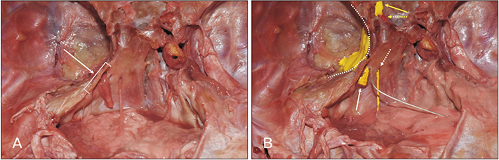
Fig. 1 (A) Ossified roof of the porus trigeminus (arrow), covering the trigeminal nerve as it enters the middle cranial fossa. The boundary of the ossification is indicated by the dashed bracket. (B) A duplicated abducens nerve dural foramen is indicated by the dissector, with one part of the nerve traveling inferomedial (dashed white arrow). The trigeminal root (solid white arrow) traverses the ossified porus trigeminus into Meckel's cave (curved dashed line). More anteriorly, the optic nerve (yellow solid arrow) and internal carotid artery (yellow dashed arrow) are colored for reference.
Reference
-
1. Burr HS, Robinson GB. An anatomical study of the Gasserian ganglion, with particular reference to the nature and extent of Meckel's cave. Anat Rec. 1925; 29:269–282.
Article2. Sabanci PA, Batay F, Civelek E, Al Mefty O, Husain M, Abdulrauf SI, Karasu A. Meckel's cave. World Neurosurg. 2011; 76:335–341.3. Wang JM, Edwards BA, Loukas M, Oskouian RJ, Tubbs RS. Supernumerary abducens nerves: a comprehensive review. World Neurosurg. 2018; 112:39–45.
Article4. Tubbs RS, Salter EG, Oakes WJ. Bony anomaly of Meckel's cave. Clin Anat. 2006; 19:75–77.
Article5. Du R, Binder DK, Halbach V, Fischbein N, Barbaro NM. Trigeminal neuralgia in a patient with a dural arteriovenous fistula in Meckel's cave: case report. Neurosurgery. 2003; 53:216–221.
Article6. Kimball D, Kimball H, Matusz P, Tubbs RS, Loukas M, Cohen-Gadol AA. Ossification of the posterior petroclinoid dural fold: a cadaveric study with neurosurgical significance. J Neurol Surg B Skull Base. 2015; 76:272–277.
Article7. Ciołkowski M, Sharifi M, Krajewski P, Ciszek B. Topography and morphometry of the porus trigeminus. Neurol Neurochir Pol. 2006; 40:173–178.8. Jannetta P. Microsurgical approach to the trigeminal nerve for tic douloureux. Prog Neurol Surg. 1976; 7:180–200.
Article9. Kiroglu Y, Calli C, Karabulut N, Oncel C. Intracranial calcifications on CT. Diagn Interv Radiol. 2010; 16:263–269.10. Bang SW, Han KR, Kim SH, Jeong WH, Kim EJ, Choi JW, Kim C. Uncommon cause of trigeminal neuralgia: tentorial ossification over trigeminal Notch. Case Rep Anesthesiol. 2015; 2015:819354.
Article11. Standefer M, Bay JW, Dohn DF. Trigeminal neuralgia secondary to a tentorial ossification: a case report. Neurosurgery. 1982; 11:527–529.
Article12. Ozveren MF, Sam B, Akdemir I, Alkan A, Tekdemir I, Deda H. Duplication of the abducens nerve at the petroclival region: an anatomic study. Neurosurgery. 2003; 52:645–652.
Article13. Tubbs RS, Radcliff V, Shoja MM, Naftel RP, Mortazavi MM, Zurada A, Loukas M, Cohen Gadol AA. Dorello canal revisited: an observation that potentially explains the frequency of abducens nerve injury after head injury. World Neurosurg. 2012; 77:119–121.
Article14. Shoja MM, Ramdhan R, Jensen CJ, Chern JJ, Oakes WJ, Tubbs RS. Embryology of the craniocervical junction and posterior cranial fossa, part I: Development of the upper vertebrae and skull. Clin Anat. 2018; 31:466–487.
Article15. Shoja MM, Ramdhan R, Jensen CJ, Chern JJ, Oakes WJ, Tubbs RS. Embryology of the craniocervical junction and posterior cranial fossa, part II: Embryogenesis of the hindbrain. Clin Anat. 2018; 31:488–500.
Article
- Full Text Links
-
- Actions
-
Cited
- CITED
-
- Close
- Share
-
- Similar articles
-
- Normal Abduction in a Patient with Duplicated Abducens Nerve
- A Case of Aberrant Abducens Nerve in a Cadaver and Review of Its Clinical Significance
- A Case of Isolated Unilateral Abducens Nerve Palsy Caused by Clival Metastasis from Rectal Cancer
- Unilateral Abducens Nerve Palsy Associated with Ruptured Anterior Communicating Artery Aneurysm
- Cystic Abducens Schwannoma without Abducens Paresis : Possible Role of Cisternal Structures in Clinical Manifestation
