Korean Circ J.
2019 Jul;49(7):642-643. 10.4070/kcj.2018.0457.
Early Transcatheter Aortic Valve Failure Accompanied with Leaflet Perforation
- Affiliations
-
- 1Division of Cardiology, Department of Internal Medicine, Yeungnam University Medical Center, Yeungnam University College of Medicine, Daegu, Korea. seranflute@gmail.com
- 2Department of Thoracic and Cardiovascular Surgery, Yeungnam University Medical Center, Yeungnam University College of Medicine, Daegu, Korea.
- KMID: 2450397
- DOI: http://doi.org/10.4070/kcj.2018.0457
Abstract
- No abstract available.
MeSH Terms
Figure
-
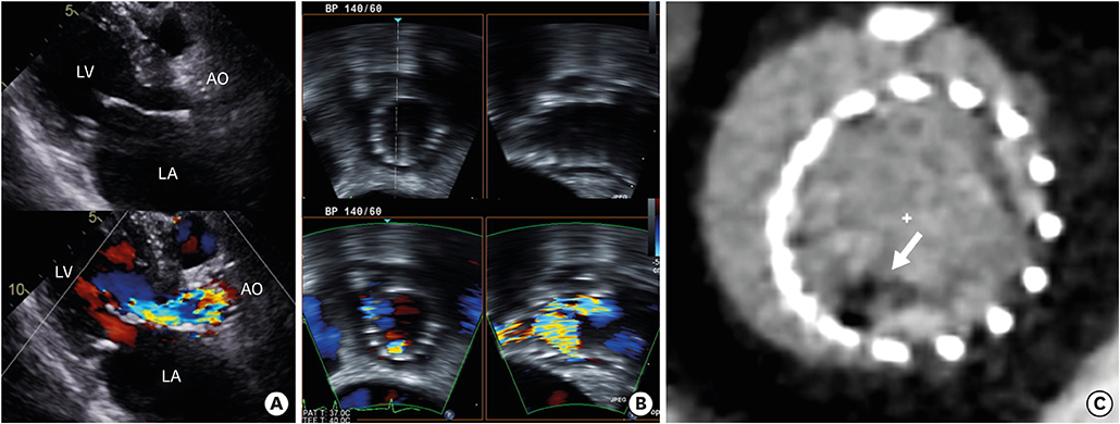
Figure 1 Pre-operative images. (A) Transthoracic echocardiography showed eccentric severe aortic regurgitation with a highly turbulent jet. (B) Transesophageal echocardiography showed eccentric severe aortic regurgitation without vegetation and abnormal leaflet thickening. (C) Computed tomography showing contrast filling defect (white arrow) within a CoreValve-prosthesis in axial view. AO = aorta; LA = left atrium; LV = left ventricle.
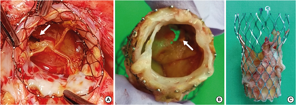
Figure 2 Intraoperative view. (A) The intraoperative view shows right coronary cusp perforation (white arrow) without vegetation and incomplete endothelization. (B) Cusp perforation can be seen from the other side. (C) Incomplete endothelization of the metallic portion of a CoreValve-prosthesis.
Reference
-
1. Mylotte D, Andalib A, Thériault-Lauzier P, et al. Transcatheter heart valve failure: a systematic review. Eur Heart J. 2015; 36:1306–1327.
Article2. Salaun E, Sportouch L, Barral PA, et al. Diagnosis of infective endocarditis after TAVR: value of a multimodality imaging approach. JACC Cardiovasc Imaging. 2018; 11:143–146.
- Full Text Links
-
- Actions
-
Cited
- CITED
-
- Close
- Share
-
- Similar articles
-
- Expanding transcatheter aortic valve replacement into uncharted indications
- A Case of Aortic Valve Endocarditis Complicated with an Aneurysm and Perforation of Anterior Mitral Leaflet
- Neo-Leaflet Failure after Comprehensive Aortic Root and Valve Reconstruction
- Anterior Mitral Leaflat Perforation in Patients with Bicuspid Aortic Valve Endocarditis
- Mitral Valve Leaflet Aneurysm-Dynamic CT and Echocardiographic Appearances
