Giant complex odontoma in the posterior mandible: A case report and literature review
- Affiliations
-
- 1Department of Oral and Maxillofacial Surgery, School of Dentistry, College of Dentistry, Wonkwang University, Iksan, Korea. omslee@wku.ac.kr
- 2Department of Oral and Maxillofacial Radiology, College of Dentistry, Wonkwang University, Iksan, Korea. siltj55@wku.ac.kr
- KMID: 2450170
- DOI: http://doi.org/10.5624/isd.2018.48.4.289
Abstract
- Odontomas are considered a type of odontogenic hamartoma, and are generally reported not to exceed 3 cm in diameter. Some authors have referred to odontomas with a diameter exceeding 3 cm as giant odontomas. As hamartomas, giant odontomas generally show no signs or symptoms, but if they perforate the mucosa to become exposed in the oral cavity, oral and maxillofacial infections can result. Surgical removal and a histopathological examination may also be required to differentiate them from osteomas, cemento-osseous dysplasia, or mixed odontogenic tumors. This report presents the case of a 28-year-old woman with a giant odontoma in the right mandibular third molar area. Based on a review of the literature published since 2010, only 11 cases of "giant" or "large" odontomas have been reported, most of which were of the complex odontoma type. It was confirmed that they tend to occur in the right posterior mandible.
Keyword
MeSH Terms
Figure
-

Fig. 1 Panoramic radiograph taken on the first visit. The radiopaque lesion can be observed around the impacted right mandibular third molar.

Fig. 2 Coronal and axial computed tomography views taken on the first visit. A. The radiopaque lesion is surrounded by a narrow radiolucent rim and dental follicle of the impacted right mandibular third molar. The radiolucent lesion of the impacted third molar is continuous with the radiolucent rim of the giant odontoma (white arrow). B. A 20×30×25 mm radiopaque lesion is observed on the right side of mandible. Expansion and thinning of the buccal cortical bone are observed on the right side of the mandible. The heterogenous radiopacity of the mass is also visible on the radiograph (black arrow).

Fig. 3 Clinical photograph showing fragments of a complex odontoma with a left mandibular impacted third molar.

Fig. 4 A postoperative panoramic image shows that the mass had been removed, the left maxillary and mandibular third molars and the right mandibular third molar had been extracted, and the left iliac block bone had been fixed with mini-plates. Maxillomandibular fixation was performed using a skeletal anchorage system screw and rubber.
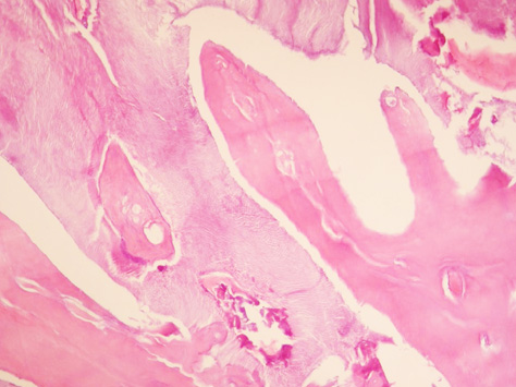
Fig. 5 Decalcified section of an odontoma shows a disorganized mass of randomly-arranged dentin intermixed with enamel matrix (hematoxylin and eosin staining, ×100).
Reference
-
1. Bagewadi SB, Kukreja R, Suma GN, Yadav B, Sharma H. Unusually large erupted complex odontoma: a rare case report. Imaging Sci Dent. 2015; 45:49–54.
Article2. Hidalgo-Sánchez O, Leco-Berrocal MI, Martínez-González JM. Metaanalysis of the epidemiology and clinical manifestations of odontomas. Med Oral Patol Oral Cir Bucal. 2008; 13:E730–E734.3. An SY, An CH, Choi KS. Odontoma: a retrospective study of 73 cases. Imaging Sci Dent. 2012; 42:77–81.
Article4. Spini PH, Spini TH, Servato JP, Faria PR, Cardoso SV, Loyola AM. Giant complex odontoma of the anterior mandible: report of case with long follow up. Braz Dent J. 2012; 23:597–600.
Article5. Perumal CJ, Mohamed A, Singh A, Noffke CE. Sequestrating giant complex odontoma: a case report and review of the literature. J Maxillofac Oral Surg. 2013; 12:480–484.
Article6. Kulkarni VK, Vanka A, Shashikiran ND. Compound odontoma associated with an unerupted rotated and dilacerated maxillary central incisor. Contemp Clin Dent. 2011; 2:218–221.
Article7. Lee J, Lee EY, Park EJ, Kim ES. An alternative treatment option for a bony defect from large odontoma using recycled demineralization at chairside. J Korean Assoc Oral Maxillofac Surg. 2015; 41:109–115.
Article8. Chrcanovic BR, Jaeger F, Freire-Maia B. Two-stage surgical removal of large complex odontoma. Oral Maxillofac Surg. 2010; 14:247–252.
Article9. Utumi ER, Cremonini CC, Pedron IG, Zambon CE, Cavalcanti MG, Ceccheti MM. Maxillary reconstruction with particulate bone graft and titanium mesh: a treatment option for large complex odontoma of the maxilla. J Dent Child (Chic). 2011; 78:124–128.10. Bodin I, Julin P, Thomsson M. Odontomas and their pathological sequels. Dentomaxillofac Radiol. 1983; 12:109–114.
Article11. Amado Cuesta S, Gargallo Albiol J, Berini Aytés L, Gay Escoda C. Review of 61 cases of odontoma. Presentation of an erupted complex odontoma. Med Oral. 2003; 8:366–373.12. Akerzoul N, Chbicheb S, El Wady W. Giant complex odontoma of mandible: a spectacular case report. Open Dent J. 2017; 11:413–419.
Article13. Baldawa RS, Khante KC, Kalburge JV, Kasat VO. Orthodontic management of an impacted maxillary incisor due to odontoma. Contemp Clin Dent. 2011; 2:37–40.
Article14. Miki Y, Oda Y, Iwaya N, Hirota M, Yamada N, Aisaki K, et al. Clinicopathological studies of odontoma in 47 patients. J Oral Sci. 1999; 41:173–176.
Article15. Vengal M, Arora H, Ghosh S, Pai KM. Large erupting complex odontoma: a case report. J Can Dent Assoc. 2007; 73:169–173.16. Ragalli CC, Ferreria JL, Blasco F. Large erupting complex odontoma. Int J Oral Maxillofac Surg. 2000; 29:373–374.
Article17. Lehman H, Lustmann J, Regev E. Removal of an extensive mandibular odontoma using an intraoral approach. Quintessence Int. 2013; 44:425–428.18. de França TR, Gueiros LA, de Castro JF, Catunda I, Leão JC, da Cruz Perez DE. Solitary peripheral osteomas of the jaws. Imaging Sci Dent. 2012; 42:99–103.
Article19. Biocic J, Macan D, Brajdic D, Manojlovic S, Butorac-Rakvin L, Hat J. Large erupting complex odontoma in a dentigerous cyst removed by a piecemeal resection. Pediatr Dent. 2010; 32:255–259.20. Mortazavi H, Baharvand M. Jaw lesions associated with impacted tooth: a radiographic diagnostic guide. Imaging Sci Dent. 2016; 46:147–157.
Article21. Dive A, Khandekar S, Bodhade A, Dhobley A. Odontoameloblastoma. J Oral Maxillofac Pathol. 2011; 15:60–64.
Article22. Reddy GS, Reddy GV, Sidhartha B, Sriharsha K, Koshy J, Sultana R. Large complex odontoma of mandible in a young boy: a rare and unusual case report. Case Rep Dent. 2014; 2014:854986.
Article23. Tomizawa M, Otsuka Y, Noda T. Clinical observations of odontomas in Japanese children: 39 cases including one recurrent case. Int J Paediatr Dent. 2005; 15:37–43.
Article
- Full Text Links
-
- Actions
-
Cited
- CITED
-
- Close
- Share
-
- Similar articles
-
- Unusually large erupted complex odontoma: A rare case report
- Ameloblastic fibro-odontoma in the mandible: a case report
- Large complex odontoma of maxillary sinus: A case report and literature review
- A Case of Complex Composite Odontoma in the Hard Palate
- A case report of ameloblastic fibro-odontioma of the mandible
