Craniometaphyseal dysplasia: Report of 2 cases with an emphasis on panoramic imaging features
- Affiliations
-
- 1Department of Oral and Maxillofacial Radiology and Dental Research Institute, School of Dentistry, Seoul National University, Seoul, Korea. hengurl@naver.com
- KMID: 2450169
- DOI: http://doi.org/10.5624/isd.2018.48.4.283
Abstract
- Craniometaphyseal dysplasia (CMD) is a rare hereditary disorder characterized by hyperostosis of the craniofacial bones and flared metaphyses of the long bones. Although some reports have described the dentomaxillofacial characteristics of CMD, including increased density of the jaw, malocclusion, and delayed eruption of the permanent teeth, only a few studies have reported the distinct imaging features of CMD on panoramic radiography. This report presents 2 cases of confirmed CMD patients with an emphasis on panoramic imaging features. The patients' images revealed hyperostosis and sclerosis of the maxilla and mandibular alveolar bone, but there was no change in the mandibular basal bone. In both cases, the mandibular condyle heads exhibited a short clubbed shape with hyperplasia of the coronoid process. For patients without clear otorhinolaryngological symptoms, common radiologic features of CMD could be visualized by routinely-taken panoramic radiographs, and further medical examinations and treatment can be recommended.
MeSH Terms
Figure
-
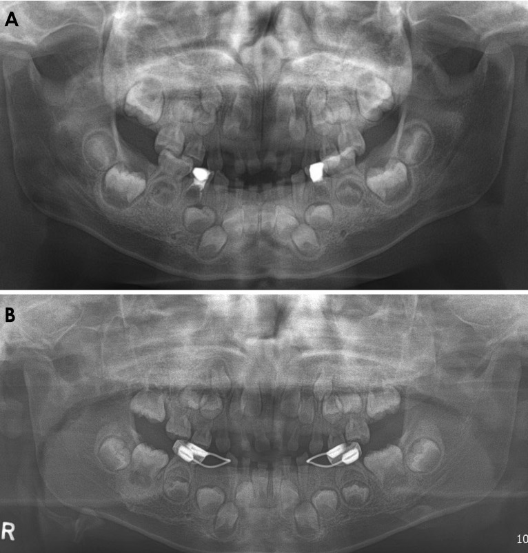
Fig. 1 A. A panoramic radiograph of the patient in case 1, showing hyperostosis and sclerosis of the alveolar bone around the developing tooth germs. The shape of the condyle was stubby and the coronoid process showed hyperplasia. The antegonial notch was underdeveloped for her age. B. A panoramic radiograph taken 2 years later. Compared with Figure 1A, sclerosis around the erupting teeth (such as the mandibular first and second molars) had increased, but sclerosis had decreased around the mandibular premolars, and the border of the bony crypts could be distinguished clearly.
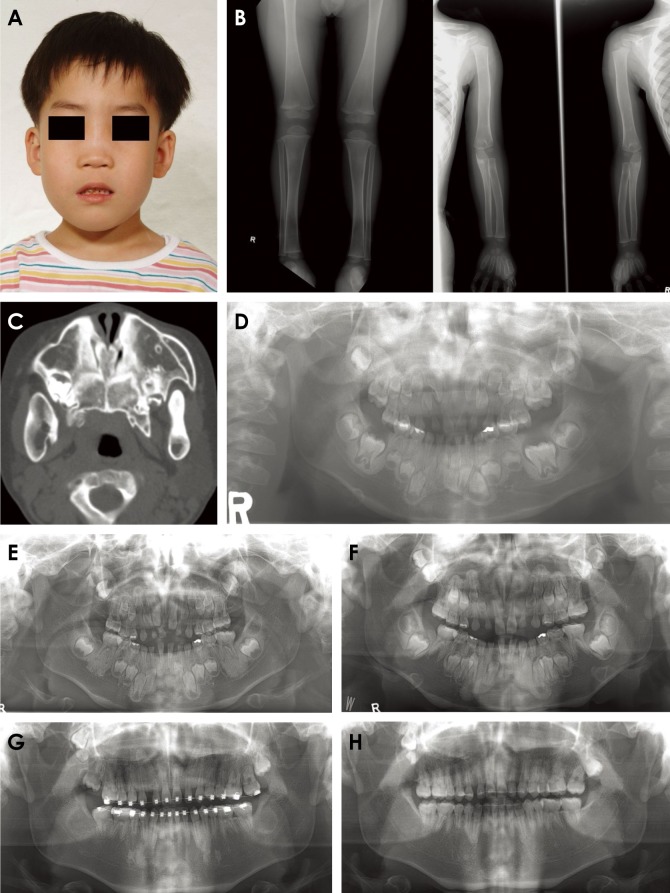
Fig. 2 A. Clinical photograph of the patient in case 2. The patient had a small nose with a wide and thickened bony nasal bridge and prominent zygomatic arches. B. Anteroposterior radiographs of the extremities, showing mild widening of the metaphysis. C. Previous computed tomography from another hospital, revealing multiple areas of hyperostosis of facial bone, including the maxilla and mandible, with bilateral obliteration of the maxillary sinus. D. A panoramic radiograph taken at the age of 6 years, showing hyperostosis and sclerotic changes in the alveolar bone around developing tooth germs. The shape of both condyles was stumpy and the antegonial notches were underdeveloped. E. A panoramic radiograph taken at the age of 8 years. All first molars had erupted, and sclerosis remained around the original tooth-bearing area. F. A panoramic radiograph taken at the age of 11 years. Sclerosis around the tooth germ area of the first molars had decreased significantly. There was increased sclerosis around the permanent canine-bearing area. G. A panoramic radiograph taken at the age of 15 years. After completion of eruption, sclerosis around the alveolar bone had decreased visibly. There was some remaining sclerosis around the canine-bearing area. H. A panoramic radiograph taken at the age of 18 years. showing no significant sclerosis around the bimaxillary alveolar bone. There were 3 impacted teeth at the bilateral posterior maxilla.
Reference
-
1. Sena Esteves S, Silva AP, Coutinho MB, Abrunhosa J, Almeida e Sousa C. Craniometaphyseal dysplasia and otolaryngology findings. Int J Pediatr Otorhinolaryngol Extra. 2015; 10:11–13.2. Dutra EH, Chen IP, Reichenberger EJ. Dental abnormalities in a mouse model for craniometaphyseal dysplasia. J Dent Res. 2013; 92:173–179. PMID: 23160629.
Article3. Thankappan S, Nair S. Craniometaphyseal dysplasia: a rare case in radiologic perspective. J Indian Acad Oral Med Radiol. 2014; 26:428–431.
Article4. Kim YH, Roh DH, Choi BY, Oh SH. Craniometaphyseal dysplasia. Acta Otolaryngol. 2005; 125:797–800. PMID: 16012045.
Article5. Twigg V, Carr S, Peres C, Mirza S. Turbinoplasty surgery for nasal obstruction in craniometaphyseal dysplasia: a case report and review of the literature. Int J Pediatr Otorhinolaryngol. 2015; 79:935–937. PMID: 25890400.
Article6. Mintz S, Velez I. Craniometaphyseal dysplasia associated with obstructive sleep apnoea syndrome. Dentomaxillofac Radiol. 2004; 33:262–266. PMID: 15533982.
Article7. Hayashibara T, Komura T, Sobue S, Ooshima T. Tooth eruption in a patient with craniometaphyseal dysplasia: case report. J Oral Pathol Med. 2000; 29:460–462. PMID: 11016689.
Article8. Lamazza L, Messina A, D’ambrosio F, Spink M, De Biase A. Craniometaphyseal dysplasia: a case report. Oral Surg Oral Med Oral Pathol Oral Radiol Endod. 2009; 107:e23–e27. PMID: 19426903.
Article9. Jackson WP, Albright F, Drewry G, Hanelin J, Rubin MI. Metaphyseal dysplasia, epiphyseal dysplasia, diaphyseal dysplasia, and related conditions. I. Familial metaphyseal dysplasia and craniometaphyseal dysplasia; their relation to leontiasis ossea and osteopetrosis; disorders of bone remodeling. AMA Arch Intern Med. 1954; 94:871–885. PMID: 13217486.10. Beighton P. Craniometaphyseal dysplasia (CMD), autosomal dominant form. J Med Genet. 1995; 32:370–374. PMID: 7616544.
Article11. Reichenberger E, Tiziani V, Watanabe S, Park L, Ueki Y, Santanna C, et al. Autosomal dominant craniometaphyseal dysplasia is caused by mutations in the transmembrane protein ANK. Am J Hum Genet. 2001; 68:1321–1326. PMID: 11326338.
Article12. Richards A, Brain C, Dillon MJ, Bailey CM. Craniometaphyseal and craniodiaphyseal dysplasia, head and neck manifestations and management. J Laryngol Otol. 1996; 110:328–338. PMID: 8733453.
Article13. Key LL Jr, Volberg F, Baron R, Anast CS. Treatment of craniometaphyseal dysplasia with calcitriol. J Pediatr. 1988; 112:583–587. PMID: 3351684.
