Imaging Sci Dent.
2018 Dec;48(4):277-281. 10.5624/isd.2018.48.4.277.
Evaluation of the relationship between sleep bruxism and pulpal calcifications in young women: A clinico-radiological study
- Affiliations
-
- 1Department of Oral and Maxillofacial Radiology, Necmettin Erbakan University, Faculty of Dentistry, Konya, Turkey. dishekmelek@gmail.com
- KMID: 2450168
- DOI: http://doi.org/10.5624/isd.2018.48.4.277
Abstract
- PURPOSE
This study was performed to investigate the relationship between sleep bruxism (SB) and pulpal calcifications in young women.
MATERIALS AND METHODS
A total of 100 female participants between 20 and 31 years of age who were referred to our radiology clinic for a dental check-up, including 59 SB and 41 non-SB patients, were sampled for the analysis. SB was diagnosed based on the American Academy of Sleep Medicine criteria. All teeth were evaluated on digital panoramic radiographs to detect pulpal calcifications, except third molars, teeth with root canal treatment, and teeth with root resorption. Binary logistic regression analysis was used to determine the risk factors for pulpal calcifications. The Spearman correlation coefficient was applied and the Pearson chi-square test was used for categorical variables. To test intra-examiner reproducibility, Cohen kappa analysis was applied. P values < .05 were considered to indicate statistical significance.
RESULTS
A total of 2800 teeth were evaluated (1652 teeth from SB patients and 1148 from non-SB patients), and 61% of patients had at least 1 dental pulpal calcification. No statistically significant relationship was found between SB and pulpal calcifications (P>0.05). In SB patients, the total number of pulpal calcifications was 129, while in non-SB patients, it was 84. Binary logistic analysis showed that SB was not a risk factor for the presence of pulpal calcifications (odds ratio, 1.19; 95% CI, 0.52-2.69, P>.05).
CONCLUSION
No relationship was found between SB and pulpal calcifications.
MeSH Terms
Figure
-
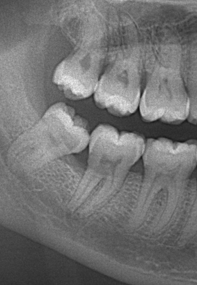
Fig. 1 Pulpal calcifications are seen in the upper and lower first and second molars on a digital panoramic image.
Reference
-
1. da Silva EJNL, Prado MC, Queiroz PM, Nejaim Y, Brasil DM, Groppo FC, et al. Assessing pulp stones by cone-beam computed tomography. Clin Oral Investig. 2017; 21:2327–2333.
Article2. Şener S, Cobankara FK, Akgünlü F. Calcifications of the pulp chamber: prevalence and implicated factors. Clin Oral Investig. 2009; 13:209–215.
Article3. Chaini K, Georgopoulou MK. General pulp calcification: literature review and case report. ENDO Endod Pract Today. 2016; 10:69–75.4. Goga R, Chandler NP, Oginni AO. Pulp stones: a review. Int Endod J. 2008; 41:457–468.
Article5. Kannan S, Kannepady SK, Muthu K, Jeevan MB, Thapasum A. Radiographic assessment of the prevalence of pulp stones in Malaysians. J Endod. 2015; 41:333–337.6. Turkal M, Tan E, Uzgur R, Hamidi MM, Çolak H, Uzgur Z. Incidence and distribution of pulp stones found in radiographic dental examination of adult Turkish dental patients. Ann Med Health Sci Res. 2013; 3:572–576.
Article7. Moody AB, Browne RM, Robinson PP. A comparison of monopolar and bipolar electrical stimuli and thermal stimuli in determining the vitality of human teeth. Arch Oral Biol. 1989; 34:701–705.
Article8. Tassoker M, Magat G, Sener S. A comparative study of cone-beam computed tomography and digital panoramic radiography for detecting pulp stones. Imaging Sci Dent. 2018; 48:201–212.
Article9. Tarim Ertas E, Inci M, Demirtas A, Ertas H, Yengil E, Sisman Y, et al. A radiographic correlation between renal and pulp stones. West Indian Med J. 2014; 63:620–625.
Article10. Giraki M, Schneider C, Schäfer R, Singh P, Franz M, Raab WH, et al. Correlation between stress, stress-coping and current sleep bruxism. Head Face Med. 2010; 6:2.
Article11. Palinkas M, De Luca Canto G, Rodrigues LA, Bataglion C, Siéssere S, Semprini M, et al. Comparative capabilities of clinical assessment, diagnostic criteria, and polysomnography in detecting sleep bruxism. J Clin Sleep Med. 2015; 11:1319–1325.
Article12. Blanco-Hungría A, Blanco-Aguilera A, Blanco-Aguilera E, Serrano-del-Rosal R, Biedma-Velázquez L, Rodriguez-Torronteras A, et al. Prevalence of the different Axis I clinical subtypes in a sample of patients with orofacial pain and temporomandibular disorders in the Andalusian Healthcare Service. Med Oral Patol Oral Cir Bucal. 2016; 21:e169–e177.13. Schmid-Schwap M, Bristela M, Kundi M, Piehslinger E. Sex-specific differences in patients with temporomandibular disorders. J Orofac Pain. 2013; 27:42–50.
Article14. Schneider C, Schaefer R, Ommerborn MA, Giraki M, Goertz A, Raab WH, et al. Maladaptive coping strategies in patients with bruxism compared to non-bruxing controls. Int J Behav Med. 2007; 14:257–261.
Article15. Landay MA, Seltzer S. The effects of excessive occlusal force on the pulp. Oral Surg Oral Med Oral Pathol. 1971; 32:623–638.
Article16. International Classification of Sleep Disorders. 3rd ed. Darien, IL: American Academy of Sleep Medicine;2014.17. Zucconi M, Ferri R. Classification of sleep disorders. In : Bassetti C, Dogas Z, Peigneux P, editors. ESRS sleep medicine textbook. Regensburg: European Sleep Research Society;2014. p. 95–109.18. Tamse A, Kaffe I, Littner MM, Shani R. Statistical evaluation of radiologic survey of pulp stones. J Endod. 1982; 8:455–458.
Article19. al-Hadi Hamasha A, Darwazeh A. Prevalence of pulp stones in Jordanian adults. Oral Surg Oral Med Oral Pathol Oral Radiol Endod. 1998; 86:730–732.20. Aslantas EE, Buzoglu HD, Karapinar SP, Cehreli ZC, Muftuoglu S, Atilla P, et al. Age-related changes in the alkaline phosphatase activity of healthy and inflamed human dental pulp. J Endod. 2016; 42:131–134.21. Sreelakhshmi , Nagaraj T, Sinha P, Goswami RD, Veerabasaviah BT. A radiographic assessment of the prevalence of idiopathic pulp calcifications in permanent teeth: a retrospective radiographic study. J Indian Acad Oral Med Radiol. 2014; 26:248–252.
Article22. Kayal RA. Distortion of digital panoramic radiographs used for implant site assessment. J Orthod Sci. 2016; 5:117–120.
Article23. Horsley SH, Beckstrom B, Clark SJ, Scheetz JP, Khan Z, Farman AG. Prevalence of carotid and pulp calcifications: a correlation using digital panoramic radiographs. Int J Comput Assist Radiol Surg. 2009; 4:169–173.
Article
- Full Text Links
-
- Actions
-
Cited
- CITED
-
- Close
- Share
-
- Similar articles
-
- Sleep and Dentistry
- Implant complications in bruxism patients
- Evaluation of the effect of two different occlusal splints on maximum occlusal force in patients with sleep bruxism: a pilot study
- Efficacy of botulinum toxin in the management of temporomandibular myofascial pain and sleep bruxism
- Relationship between self-reported bruxism and torus mandibularis of patients with temporomandibular disorders
