Normal Korean Femoral Neck Anteversion, Acetabular Anteversion and Combined Anteversion Measured with Computed Tomography
- Affiliations
-
- 1Department of Orthopedic Surgery, Cheju Halla General Hospital, Jeju, Korea. ulsanos@naver.com
- KMID: 2450146
- DOI: http://doi.org/10.4055/jkoa.2019.54.3.261
Abstract
- PURPOSE
The authors measured the anteversion of the femoral neck and acetabulum and the sum of the two values in normal Korean people by computed tomography. The authors examined the normal range of the values to analyze the difference in sex and sides as well as the relationship between the femoral neck and acetabular anteversion.
MATERIALS AND METHODS
The authors measured the anteversion of the femoral neck and acetabulum in 118 normal Korean adult males and 114 females aged between 21 and 49 on both the right and left sides by computed tomography and calculated the sum of anteversion. The authors analyzed the mean and standard deviation, and investigated the sex differences and side differences, as well as the relationship between the acetabular anteversion and femoral neck anteversion statistically.
RESULTS
The anteversion of the acetabulum in males was 15.3°±6.1° on the right side and 15.3°±6.6° on the left side. The anteversion of the femoral neck in males was 5.3°±7.6° on the right side and 1.5°±9.2° on the left side according to the Hernandez et al. method. The anteversion of acetabulum in females was 16.8°±5.4° on the right side and 16.3°±5.8° on the left side. The anteversion of femoral neck in females was 10.3°±8.2° on the right side and 7.9°±8.2° on the left side according to Hernandez et al. method. No difference in acetabular anteversion, and a significant difference in the femoral neck anteversion on both the right and left sides were observed between males and females. No difference of acetabular anteversion was observed between the right and left sides, but a significant difference in femoral neck anteversion was noted between the right and left sides measured by either the Hernandez et al. method or Weiner et al. method. The Pearson coefficient revealed no correlation between the femoral neck anteversion and acetabular anteversion.
CONCLUSION
No difference in the acetabular anteversion was observed, but there was a significant difference in femoral neck anteversion between males and females. A significant difference in femoral neck anteversion was observed between the right and left sides. No correlation was noted between the anteversion of the femoral neck and acetabulum.
Keyword
Figure
-
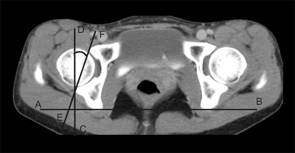
Figure 1. Method of measuring the acetabular anteversion. Line AB is drawn by connecting the most posterior point of the posterior column of the acetabulum. Line CD is perpendicular to line AB. Line EF is drawn by connecting the anterior and posterior acetabular edge. The angle between line CD and line EF is the acetabular anteversion.
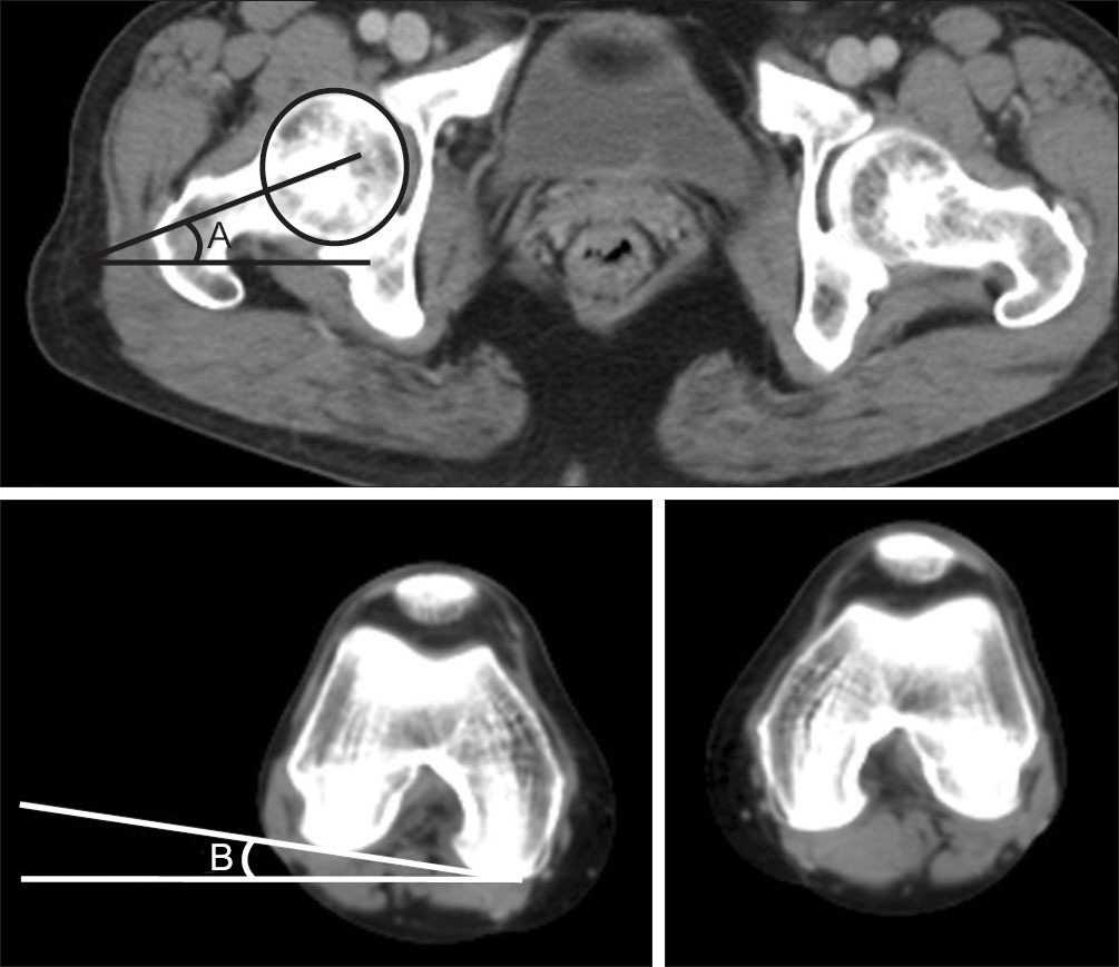
Figure 2. Method of measuring the femoral neck anteversion. A is the angle between the horizontal line and the line of the femoral neck axis, B is the angle between the horizontal line and the line connecting the posterior surface of the distal femoral medial condyle and lateral condyle. A minus B is the femoral neck anteversion.
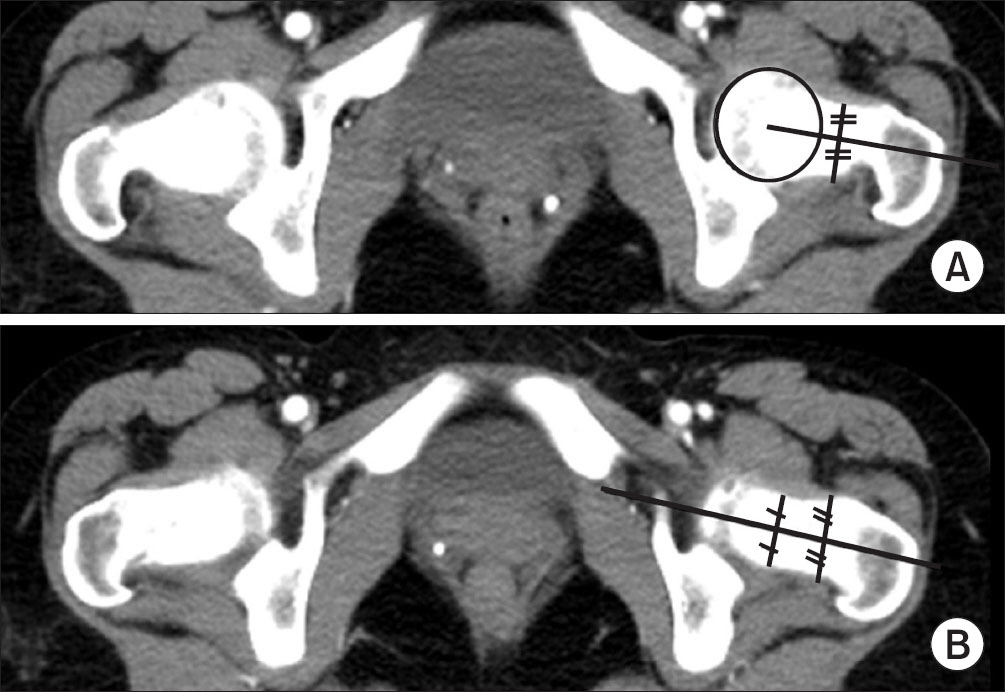
Figure 3. Defining the axis of femoral neck: (A) The Hernandez et al. method9) is defining the femoral neck axis as the line which connects the center of the femoral head and that of the isthmus of the neck. (B) The Weiner et al. method10) is defining the femoral neck axis as the midline between the ventral and dorsal cortices of the femoral neck.
Reference
-
References
1. Mckibbin B. Anatomical factors in the stability of the hip joint in the newborn. J Bone Joint Surg Br. 1970. 52:148–59.
Article2. Tönnis D, Heinecke A. Acetabular and femoral anteversion: relationship with osteoarthritis of the hip. J Bone Joint Surg Am. 1999. 81:1747–70.3. Reikerås O, Bjerkreim I, Kolbenstvedt A. Anteversion of the acetabulum and femoral neck in normals and in patients with osteoarthritis of the hip. Acta Orthop Scand. 1983. 54:18–23.4. Sugano N, Noble PC, Kamaric E. A comparison of alternative methods of measuring femoral anteversion. J Comput Assist Tomogr. 1998. 22:610–4.
Article5. Murphy SB, Simon SR, Kijewski PK, Wilkinson RH, Griscom NT. Femoral anteversion. J Bone Joint Surg Am. 1987. 69:1169–76.
Article6. Murray DW. The definition and measurement of acetabular orientation. J Bone Joint Surg Br. 1993. 75:228–32.
Article7. Yao L, Yao J, Gold RH. Measurement of acetabular version on the axiolateral radiograph. Clin Orthop Relat Res. 1995. 316:106–11.
Article8. Jain AK, Maheshwari AV, Singh MP, Nath S, Bhargava SK. Femoral neck anteversion: a comprehensive Indian study. Indian J Orthop. 2005. 39:137–44.
Article9. Hernandez RJ, Tachdjian MO, Poznanski AK, Dias LS. CT determination of femoral torsion. AJR Am J Roentgenol. 1981. 137:97–101.
Article10. Weiner DS, Cook AJ, Hoyt WA Jr, Oravec CE. Computed tomography in the measurement of femoral anteversion. Orthopedics. 1978. 1:299–306.
Article11. Maruyama M, Feinberg JR, Capello WN, D’Antonio JA. The Frank Stinchfield Award: morphologic features of the acetabulum and femur: anteversion angle and implant positioning. Clin Orthop Relat Res. 2001. 393:52–65.12. Vandenbussche E, Saffarini M, Delogé N, Moctezuma JL, Nogler M. Hemispheric cups do not reproduce acetabular rim morphology. Acta Orthop. 2007. 78:327–32.
Article13. Köhnlein W, Ganz R, Impellizzeri FM, Leunig M. Acetabular morphology: implications for joint-preserving surgery. Clin Orthop Relat Res. 2009. 467:682–91.
Article14. Zeng Y, Wang Y, Zhu Z, Tang T, Dai K, Qiu S. Differences in acetabular morphology related to side and sex in a Chinese population. J Anat. 2012. 220:256–62.
Article15. Chang JS, Park JH, Shon HC. . Incidence of acetabular retroversion in dysplastic hip. J Korean Orthop Assoc. 2004. 39:675–8.
Article16. Jamali AA, Mladenov K, Meyer DC. . Anteroposterior pelvic radiographs to assess acetabular retroversion: high validity of the "cross-over-sign". J Orthop Res. 2007. 25:758–65.
Article17. Zeng WN, Wang FY, Chen C. . Investigation of association between hip morphology and prevalence of osteoarthritis. Sci Rep. 2016. 6:23477.
Article18. Khang G, Choi K, Kim CS, Yang JS, Bae TS. A study of Korean femoral geometry. Clin Orthop Relat Res. 2003. 406:116–22.
Article19. Toogood PA, Skalak A, Cooperman DR. Proximal femoral anatomy in the normal human population. Clin Orthop Relat Res. 2009. 467:876–85.
Article20. Lavy CB, Msamati BC, Igbigbi PS. Racial and gender variations in adult hip morphology. Int Orthop. 2003. 27:331–3.
Article21. Hoaglund FT, Low WD. Anatomy of the femoral neck and head, with comparative data from Caucasians and Hong Kong Chinese. Clin Orthop Relat Res. 1980. 152:10–6.
Article22. Van Houcke J, Yau WP, Yan CH. . Prevalence of radiographic parameters predisposing to femoroacetabular impingement in young asymptomatic Chinese and white subjects. J Bone Joint Surg Am. 2015. 97:310–7.23. Fabricant PD, Fields KG, Taylor SA, Magennis E, Bedi A, Kelly BT. The effect of femoral and acetabular version on clinical outcomes after arthroscopic femoroacetabular impingement surgery. J Bone Joint Surg Am. 2015. 97:537–43.
Article24. Han CD, Lee YH, Yang KH. . Predicting proximal femur rotation by morphological analyses using translucent 3-dimensional computed tomography. Arch Orthop Trauma Surg. 2012. 132:1747–52.
Article25. Dorr LD, Malik A, Dastane M, Wan Z. Combined anteversion technique for total hip arthroplasty. Clin Orthop Relat Res. 2009. 467:119–27.
Article26. Amuwa C, Dorr LD. The combined anteversion technique for acetabular component anteversion. J Arthroplasty. 2008. 23:1068–70.
Article
- Full Text Links
-
- Actions
-
Cited
- CITED
-
- Close
- Share
-
- Similar articles
-
- Usefulness of a Modular Hip System for Combined Anteversion in Cementless Total Hip Arthroplasty
- Relationship between Femoral Anteversion and Tibial Torsion in Intoeing Gait
- Measurement of Femoral Anteversion in the PACS Image Viewer
- Computed Tomography in Evaluation of Femoral Anteversion: Transverse Section Versus Axial Oblique Section
- Increase of Femoral Anteversion after Experimental Induction of Avascular Necrosis of Femoral Head
