A Case of Squamous Cell Carcinoma of Cervical Esophagus with Metastasis to Thyroid Gland
- Affiliations
-
- 1Department of Otorhinolaryngology-Head & Neck Surgery, Korea Cancer Center Hospital, KIRAMS, Seoul, Korea. ijchoiorl@gmail.com
- KMID: 2449068
- DOI: http://doi.org/10.11106/ijt.2019.12.1.64
Abstract
- Despite thyroid is a highly vascularized organ, clinically significant metastatic spread to the thyroid is considered uncommon. There is a reported incidence of up to 24.0% metastases to the thyroid in autopsy series. The most frequently noted primary sites are the kidney, breast, and lung. The metastatic spread of alimentary tract is quite rare, and the majority comes from the colo-rectum. We present a case of squamous cell carcinoma of the cervical esophagus presenting as thyroid nodule in an apparently healthy 54 year-old male patient. This might be the first case of esophageal carcinoma metastases to the thyroid presenting in South Korea.
MeSH Terms
Figure
-
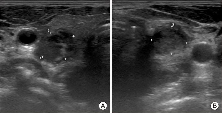
Fig. 1 Thyroid US shows 1.3×1.1×2.2 cm sized thyroid nodule with hypoechoic irregular margin in right thyroid gland (A). At the same time, 1.2×1.0×1.3 cm sized hypoechoic irregular margin nodule in left thyroid gland (B). Sono-guided FNAB was performed in these nodules.
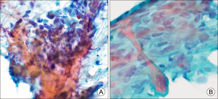
Fig. 2 Fine needle aspiration cytology smear (A) from right thyroid nodule and (B) from level IV lymph node. High power view demonstrates sheets of atypical polygonal cells with prominent nucleoli in necrotic background. Some cells have tadpole appearance with keratinization (Papanicolaou stain, ×400).
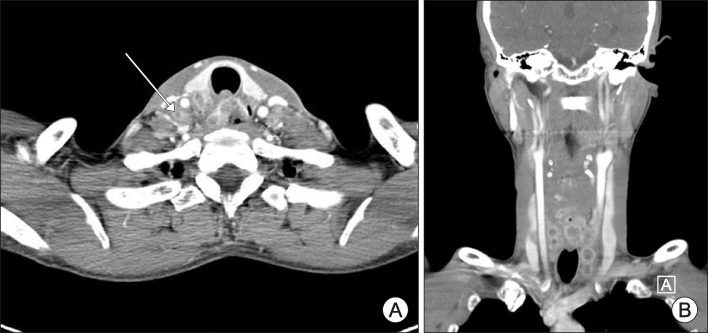
Fig. 3 Axial (A) and coronal (B) images from a contrast-enhanced computed tomography of the thyroid show multiple conglomerated necrotic lymphadenopathies in central neck, both lateral neck, and mediastinum with posterior aspect of thyroid gland (long arrow) and bilateral tracheoesophageal groove area involvement.
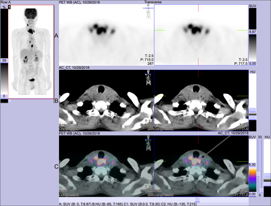
Fig. 4 PET/CT images show high focal 18F-FDG uptake in the cervical esophagus and the posterior aspect of both thyroid glands (maximum SUV=16.2), high focal uptakes in right lateral neck LNs (maximum SUV=6.9), and several focal uptakes in upper thoracic esophagus (long arrow) (two lesions; maximum SUV=4.6 and 4.4) and mid thoracic esophagus (two lesions; maximum SUV=11.4 and 3.0).
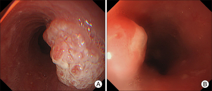
Fig. 5 Endoscopic appearance shows a round protruding polyp measuring 3.0 cm, located at the of the point 32 cm far from the upper esophageal sphincter and an ulcerofungating mass located at the of the point 22 cm far from the upper esophageal sphincter (A). And an ulcerofungating mass located at the of the point 22 cm far from the upper esophageal sphincter (B).
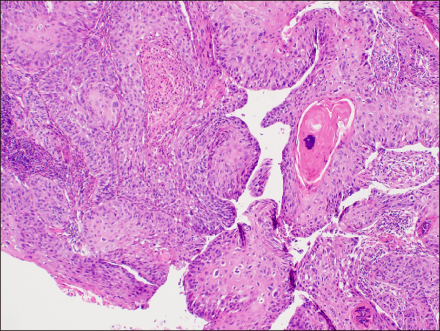
Fig. 6 Invasive squamous cell carcinoma in endoscopic biopsy specimen from upper esophagus. Moderately differentiated lesions show sheets of large polygonal malignant cells containing keratin formation (Hematoxylin & Eosin stain, ×100).
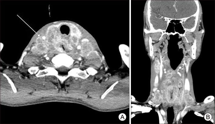
Fig. 7 1 month later follow up thyroid CT. Axial (A) and coronal (B) images show markedly increasing in size and number of conglomerated metastatic lymphadenopathies in both retropharyngeal space, central neck, both lateral neck (long arrow) and mediastinum with invasion of both sides of tracheoesophageal groove, both sides of thyroid gland and cricoid cartilage.
Reference
-
1. Lam KY, Lo CY. Metastatic tumors of the thyroid gland: a study of 79 cases in Chinese patients. Arch Pathol Lab Med. 1998; 122(1):37–41.2. Nakhjavani MK, Gharib H, Goellner JR, van Heerden JA. Metastasis to the thyroid gland. A report of 43 cases. Cancer. 1997; 79(3):574–578.3. Lin JD, Weng HF, Ho YS. Clinical and pathological characteristics of secondary thyroid cancer. Thyroid. 1998; 8(2):149–153.
Article4. Pillay SP, Angorn IB, Baker LW. Tumour metastasis to the thyroid gland. S Afr Med J. 1977; 51(15):509–512.5. Ericsson M, Biorklund A, Cederquist E, Ingemansson S, Akerman M. Surgical treatment of metastatic disease in the thyroid gland. J Surg Oncol. 1981; 17(1):15–23.
Article6. Czech JM, Lichtor TR, Carney JA, van Heerden JA. Neoplasms metastatic to the thyroid gland. Surg Gynecol Obstet. 1982; 155(4):503–505.7. McCabe DP, Farrar WB, Petkov TM, Finkelmeier W, O'Dwyer P, James A. Clinical and pathologic correlations in disease metastatic to the thyroid gland. Am J Surg. 1985; 150(4):519–523.
Article8. Yoshida A, Imamura A, Tanaka H, Hirano M, Kamma H, Ueno E, et al. A case of metastasis from gastric cancer to the thyroid gland. Jpn J Surg. 1989; 19(4):480–484.
Article9. Wychulis AR, Beahrs OH, Woolner LB. Metastasis of carcinoma to the thyroid gland. Ann Surg. 1964; 160:169–177.
Article10. Cumbo-Nacheli G, de Sanctis JT, Chung MH. Proximal esophageal adenocarcinoma presenting as a thyroid mass: case report and review of the literature. Thyroid. 2007; 17(3):267–269.
Article11. Reese J, Chebolu A, Shen Y, Mihlon F. Case report: Diffuse metastatic infiltration of the thyroid by esophageal adenocarcinoma mimicking non-neoplastic thyroid disease. Radiol Case Rep. 2018; 13(1):108–111.
Article12. Shaheen O, Ghibour A, Alsaid B. Esophageal cancer metastases to unexpected sites: A systematic review. Gastroenterol Res Pract. 2017; 2017:1657310.
Article13. Gooptu S, Sharma S, Singh G, Ali I. Uncommon metastasis to thyroid gland presenting as a thyroid nodule. IJCRI. 2013; 4(11):615–618.
Article14. Basu S, Nair N, Borges AM. Squamous cell carcinoma of esophagus masquerading as solitary thyroid nodule. Indian J Cancer. 2005; 42(4):205–207.15. Ahn D, Sohn JH. Anaplastic thyroid carcinoma: Experience of a single institute. Korean J Otorhinolaryngol-Head Neck Surg. 2012; 55(1):37–41.
Article16. Cook AM, Vini L, Harmer C. Squamous cell carcinoma of the thyroid: outcome of treatment in 16 patients. Eur J Surg Oncol. 1999; 25(6):606–609.
Article17. Kumar PV, Malekhusseini SA, Talei AR. Primary squamous cell carcinoma of the thyroid diagnosed by fine needle aspiration cytology. A report of two cases. Acta Cytol. 1999; 43(4):659–662.
Article18. Chen H, Nicol TL, Udelsman R. Clinically significant, isolated metastatic disease to the thyroid gland. World J Surg. 1999; 23(2):177–180. discussion 81.
Article19. Mirallie E, Rigaud J, Mathonnet M, Gibelin H, Regenet N, Hamy A, et al. Management and prognosis of metastases to the thyroid gland. J Am Coll Surg. 2005; 200(2):203–207.
Article20. Chen ED, Cheng P, Yan XQ, Ye YL, Chen CZ, Ji XH, et al. Metastasis of distal esophageal carcinoma to the thyroid with presentation simulating primary thyroid carcinoma: a case report and review of the literature. World J Surg Oncol. 2014; 12:106.
Article21. Murakami S, Yashuda S, Nakamura T, Mishima Y, Iida H, Okano H, et al. A case of renal cell carcinoma with metastasis to the thyroid gland and concomitant early gastric cancer. Surg Today. 1993; 23(2):153–158.
Article
- Full Text Links
-
- Actions
-
Cited
- CITED
-
- Close
- Share
-
- Similar articles
-
- Synchronous thyroid carcinoma and squamous cell carcinoma: A case report
- Basaloid-Squamous Carcinoma of the Esophagus: A case report
- Intrathyroidal metastasis of tonsillar squamous cell carcinoma masquerading as a primary thyroid tumor
- Fine Needle Aspiration Cytology of Squamous Cell Carcinoma of the Thyroid: Report of A Case
- A Case of Primary Squamous Cell Carcinoma of the Thyroid Gland
