A Case of Cholesteatoma of Maxillary Sinus
- Affiliations
-
- 1Department of Otolaryngology-Head and Neck Surgery, School of Medicine, Catholic University of Daegu, Daegu, Korea. miky@cu.ac.kr
- KMID: 2449028
- DOI: http://doi.org/10.18787/jr.2019.26.1.43
Abstract
- Cholesteatoma is common disease entity within the middle ear cavity but is rarely found in the paranasal sinuses, especially the maxillary sinus. We experienced a case of cholesteatoma of the maxillary sinus without history of previous trauma or operation. The patient was not improved by functional endoscopic sinus surgery. The mucosa of the maxillary sinus was removed through the Caldwell-Luc approach, and heavy saline irrigation was performed. After reoperation, the postoperative period was uneventful, and there was no sign of recurrence on endoscopic examination.
Keyword
MeSH Terms
Figure
-
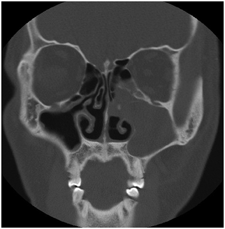
Fig. 1 Preoperative PNS CT showing non-homogeneous soft tissue density in the left maxillary sinus without bony remodeling.
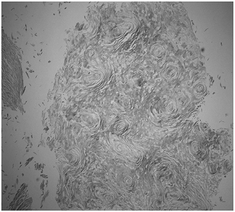
Fig. 2 Histopathological examination of specimen from maxillary sinus; Keratinous material (Hematoxylin and Eosin ×40).
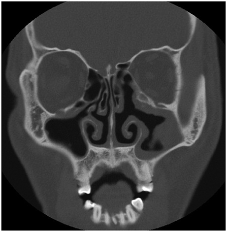
Fig. 3 PNS CT finding after 1 year of endoscopic sinus surgery showing mucosal thickening in left maxillary sinus without bony remodeling.
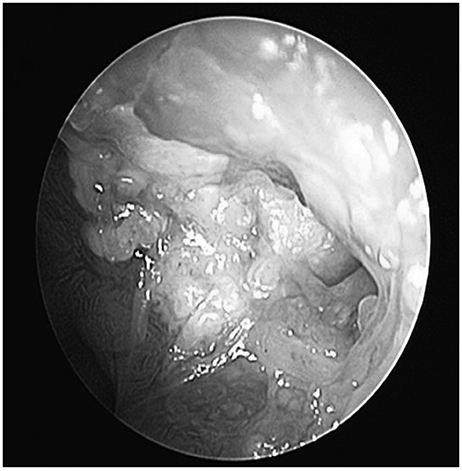
Fig. 4 Endoscopic finding of left maxillary sinus opening showing whitish keratin material.
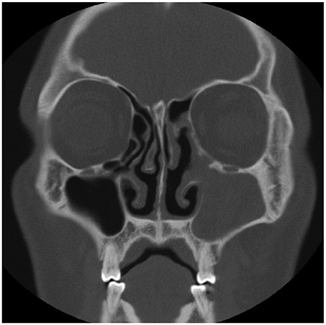
Fig. 5 PNS CT finding after 3 years of endoscopic sinus surgery showing worsened soft tissue density in maxillary sinus without bony remodeling.
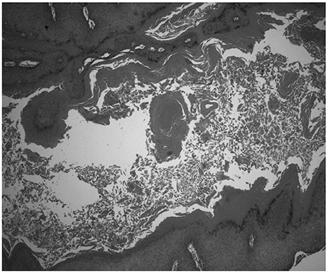
Fig. 6 Histopathological finding of maxillary sinus mucosa; fully differentiated, stratified squamous epithelium with lamellar sheets of keratin materials on connective tissue infiltrated with inflammatory cell (Hematoxylin and Eosin ×40).
Reference
-
1. Hammami B, Mnejja A, Chakroun A, Achour I, Chakroun A, Charfeddine I, et al. Cholesteatoma of the frontal sinus. Eur Ann Otorhinolaryngol Head Neck Dis. 2010; 127:213–216.
Article2. Park SH, Baek SH, Song TH, Cha YJ. Cholesteatoma of the maxillary sinus. Korean J Otolaryngol-Head Neck Surg. 1999; 42:522–525.3. Puttamadaiah GM, Vijayashree MS, Viswanatha BO, Kaur JA. Cholesteatoma of maxillary sinus mimicking malignancy. Research in Otolaryngology. 2014; 3(4):57–59.4. Min HJ, Shin JH, Kim KS. Cholesteatoma of maxillary sinus: What is the best surgical approach? J Craniofac Surg. 2016; 27:963–966.5. Viswanatha B, Nayah L, Karthik S. Cholesteatoma of the maxillary sinus. Ear Nose Throat J. 2007; 86:351–353.
Article6. Hansen S, Sorensen CH, Stage J, Mouritzen A, Cayé-Thomasen P. Massive cholesteatoma of the frontal sinus: case report and review of the literature. Auris Nasus Larynx. 2007; 34:387–392.
Article7. Hopp ML, Montgomery WW. Primary and secondary keratomas of the frontal sinus. Laryngoscope. 1984; 94:628–632.
Article8. Viswanatha BO, Nayak KR, Karthik SH. Cholesteatoma of the maxillary sinus. Ear Nose Throat J. 2007; 86:351–353.
Article9. Lee JM, Ryu NG, Choi IS. A case of maxillary sinus cholesteatoma originating from the retromaxillary sinus wall. Int J Otolaryngol. 2015; 4:325–328.
Article
- Full Text Links
-
- Actions
-
Cited
- CITED
-
- Close
- Share
-
- Similar articles
-
- Cholesteatoma of the Maxillary Sinus
- A Case of Actinomycosis of the Maxillary Sinus Accompanied with Fungal Ball
- Delayed Occurrence of Maxillary Sinusitis after Simultaneous Maxillary Sinus Augmentation and Implant: A Case Report and Literature Review
- Organized Hematoma Presenting with Periorbital Swelling: A Case Report and Review of Literatures
- A Postoperative Cheek Cyst Originating from the Maxillary Sinus with Orbital Involvement
