Ann Rehabil Med.
2019 Apr;43(2):230-233. 10.5535/arm.2019.43.2.230.
Fahr's Disease With Intracerebral Hemorrhage at the Uncommon Location: A Case Report
- Affiliations
-
- 1Department of Physical Medicine and Rehabilitation, Ulsan University Hospital, Ulsan, Korea. crkim@uuh.ulsan.kr
- KMID: 2449006
- DOI: http://doi.org/10.5535/arm.2019.43.2.230
Abstract
- Fahr's disease (FD) is a rare neurologic disorder characterized by the symmetric and bilateral intracerebral calcification in a patient. We describe the case of a 65-year-old woman who presented with gait disturbance, abnormal mentality, and visual field defect. The result of a brain computerized tomography showed spontaneous intracranial hemorrhage in the right parieto-occipital area, and also showed the incidence of symmetric and bilateral intracerebral calcification. Moreover, laboratory studies indicated characteristic hypoparathyroidism. This brings us to understand that additionally, one of her sons also presented with similar intracerebral calcification, and was subsequently diagnosed with FD. Thus, her case was consistent with that of a patient experiencing FD. The patient had hypertension, which we now know might have caused the intracerebral hemorrhage. However, this patient's brain lesions were in uncommon locations for spontaneous intracerebral hemorrhage, and the lesions were noted as occurring away from the identified heavily calcified areas. Thus, it seemed that the massive calcification of cerebral vessels in the basal ganglia, the most common site of intracerebral hemorrhage, might have prevented a hypertensive intracerebral hemorrhage. Eventually, an intracerebral hemorrhage occurred in an uncommon location in the patient's brain.
MeSH Terms
Figure
-

Fig. 1. Computed tomography scan of the patient’s brain shows an intracerebral hemorrhage in the right parietooccipital area, with calcification of bilateral periventricular white matter, thalamus, and cerebellum.
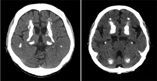
Fig. 2. Computed tomography scan of the younger son’s brain shows a bilateral basal ganglia, thalamus and cerebellar calcification.
Reference
-
1. Fahr KT. Idiopathische verkalkung der hirngefasse. Zentralbl Allg Pathol. 1930; 50:129–33.2. Manyam BV. What is and what is not ‘Fahr’s disease’. Parkinsonism Relat Disord. 2005; 11:73–80.
Article3. Wszolek ZK, Baba Y, Mackenzie IR, Uitti RJ, Strongosky AJ, Broderick DF, et al. Autosomal dominant dystoniaplus with cerebral calcifications. Neurology. 2006; 67:620–5.
Article4. Swami A, Kar G. Intracranial hemorrhage revealing pseudohypoparathyroidism as a cause of fahr syndrome. Case Rep Neurol Med. 2011; 2011:407567.
Article5. Al-Jehani H, Ajlan A, Sinclair D. Fahr’s disease presenting with aneurysmal subarachnoid hemorrhage. J Clin Imaging Sci. 2012; 2:27.
Article6. Sgulo FG, di Nuzzo G, de Notaris M, Seneca V, Catapano G. Cerebrovascular disorders and Fahr’s disease: report of two cases and literature review. J Clin Neurosci. 2018; 50:163–4.7. Baba Y, Broderick DF, Uitti RJ, Hutton ML, Wszolek ZK. Heredofamilial brain calcinosis syndrome. Mayo Clin Proc. 2005; 80:641–51.
Article8. Saleem S, Aslam HM, Anwar M, Anwar S, Saleem M, Saleem A, et al. Fahr’s syndrome: literature review of current evidence. Orphanet J Rare Dis. 2013; 8:156.
Article9. Chalkias SM, Magnaldi S, Cova MA, Longo R, Pozzi-Mucelli RS. Fahr disease: significance and predictive value of CT and MR findings. Eur Radiol. 1992; 2:570–5.
Article10. Zafar A, Khan FS. Clinical and radiological features of intracerebral haemorrhage in hypertensive patients. J Pak Med Assoc. 2008; 58:356–8.
- Full Text Links
-
- Actions
-
Cited
- CITED
-
- Close
- Share
-
- Similar articles
-
- A Case of Fahr's Disease Presenting with Frontal Lobe Dysfunction
- Epileptic Seizure Revealing a Fahr's Syndrome with Pseudohypoparathyroidism: A Case Report
- Cerebrovascular Disease during Pregnancy
- Mirror-Image Intracerebral Hemorrhage in Basal Ganglia: Case Report
- A Case of Cerebral Amyloid Angiopathy-related Intracerebral Hemorrhage
