Comparison of Skyline View and Intraoperative Computed Tomography for Detecting Protruded Screws during Volar Plating of Distal Radius Fractures
- Affiliations
-
- 1Department of Orthopedic Surgery, Wonkwang University School of Medicine, Iksan, Korea. kanghongje@hanmail.net
- KMID: 2448044
- DOI: http://doi.org/10.12790/ahm.2019.24.2.112
Abstract
- PURPOSE
The aim of our study was to compare and analyze intraoperative fluoroscopy (skyline view) and mobile cone-beam computed tomography (CBCT) for detecting protruded screws in volar locked plating used for distal radius fractures.
METHODS
We carried out a prospective analysis of 35 patients who had undergone both intraoperative fluoroscopy and mobile CBCT. The patients had all undergone volar locking plate fixation for a distal radial fracture at our institution between January and May 2017. Skyline view and mobile CBCT were carried out and protruded screws were replaced. Screw tip cortex distance (STCD) was measured using skyline view and mobile CBCT and compared with each area of the distal radius.
RESULTS
Three screws were found to be protruding after skyline view, and further seven screws were found to be protruding after computed tomography (CT) scan. The mean STCD for each compartment was 3.8±0.6 mm, 3.5±1.8 mm, 2.2±1.3 mm, 3.7±1.6 mm, and 3.9±1.4 mm in the skyline view, respectively, and 3.5±0.7 mm, 0.8±1.6 mm, 0.9±1.1 mm, 2.1±1.6 mm, and 3.7±1.9 mm in the CT scan, respectively (p<0.05). The mean STCD of all screws was 1.2 mm longer in the skyline view than in the CT scan.
CONCLUSION
The skyline view showed approximately 1-2 mm difference compared to CBCT; therefore, it would be better to insert the screw 2 mm shorter than seen in the skyline view.
MeSH Terms
Figure
-
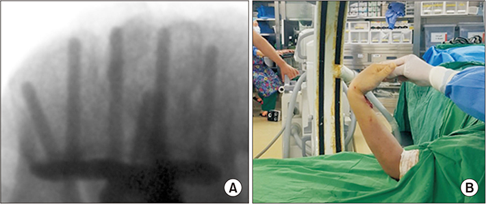
Fig. 1 (A) Skyline view of the distal radius. (B) Skyline view was taken on the C-arm with wrist flexion of 75°.
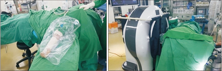
Fig. 2 Mobile cone-beam computed tomography (CBCT) (Phion™; NanoFocusRay Co., Ltd.) available in the operating room. Mobile CBCT has its own shielding structure so that it prevents radiation exposure to other part of the body.
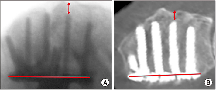
Fig. 3 The distance between the tip of the screw tip and dorsal cortex (STCD) was measured and compared for each compartment. (A) Skyline view. (B) Mobile cone-beam computed tomography. Up down arrows: STCD.
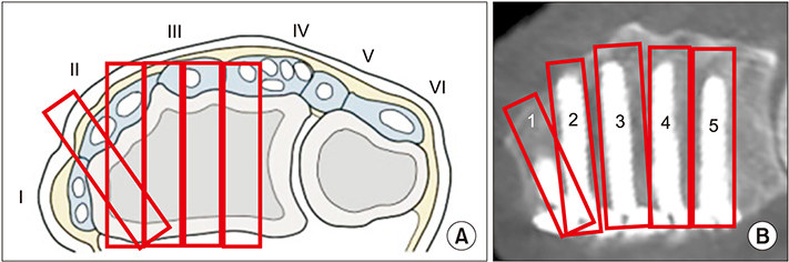
Fig. 4 (A) Extensor compartment of radial dorsal cortex. (B) The dorsal cortex of radius was divided into five areas (1, 2, 3, 4, 5) and mean screw tip cortex distance was compared for each compartment.
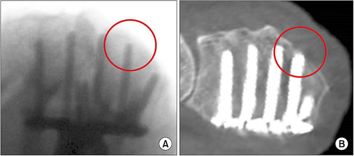
Fig. 5 Comparison of screw tip cortex distance measured by skyline view and mobile cone-beam computed tomography (CBCT). The screw in the circle is not protruded in skyline view, whereas it is protruded in the mobile CBCT. (A) Skyline view. (B) Mobile CBCT.
Reference
-
1. Downing ND, Karantana A. A revolution in the management of fractures of the distal radius? J Bone Joint Surg Br. 2008; 90:1271–1275.
Article2. Al-Rashid M, Theivendran K, Craigen MA. Delayed ruptures of the extensor tendon secondary to the use of volar locking compression plates for distal radial fractures. J Bone Joint Surg Br. 2006; 88:1610–1612.
Article3. Arora R, Lutz M, Hennerbichler A, Krappinger D, Espen D, Gabl M. Complications following internal fixation of unstable distal radius fracture with a palmar locking-plate. J Orthop Trauma. 2007; 21:316–322.
Article4. Benson EC, DeCarvalho A, Mikola EA, Veitch JM, Moneim MS. Two potential causes of EPL rupture after distal radius volar plate fixation. Clin Orthop Relat Res. 2006; 451:218–222.
Article5. Kang JW, Park JW. Complications of distal radius fracture. J Korean Orthop Assoc. 2013; 48:165–174.
Article6. Tarallo L, Mugnai R, Zambianchi F, Adani R, Catani F. Volar plate fixation for the treatment of distal radius fractures: analysis of adverse events. J Orthop Trauma. 2013; 27:740–745.7. Rhee PC, Dennison DG, Kakar S. Avoiding and treating perioperative complications of distal radius fractures. Hand Clin. 2012; 28:185–198.
Article8. Ozer K, Wolf JM, Watkins B, Hak DJ. Comparison of 4 fluoroscopic views for dorsal cortex screw penetration after volar plating of the distal radius. J Hand Surg Am. 2012; 37:963–967.
Article9. Ozer K, Toker S. Dorsal tangential view of the wrist to detect screw penetration to the dorsal cortex of the distal radius after volar fixed-angle plating. Hand (NY). 2011; 6:190–193.
Article10. Granville-Chapman J, Hacker AG, Keightley A, Sarkhel T, Monk J, Gupsta R. The distal radius skyline view: validation of a new technique for determining dorsal screw penetration in locked volar plating of the distal radius. Orthop Proc. 2012; 94:(SUPP_XXXII). 16.11. Riddick AP, Hickey B, White SP. Accuracy of the skyline view for detecting dorsal cortical penetration during volar distal radius fixation. J Hand Surg Eur Vol. 2012; 37:407–411.
Article12. Ganesh D, Service B, Zirgibel B, Koval K. The detection of prominent hardware in volar locked plating of distal radius fractures: intraoperative fluoroscopy versus computed tomography. J Orthop Trauma. 2016; 30:618–621.
Article13. Vaiss L, Ichihara S, Hendriks S, Taleb C, Liverneaux P, Facca S. The utility of the fluoroscopic skyline view during volar locking plate fixation of distal radius fractures. J Wrist Surg. 2014; 3:245–249.
Article14. McKay SD, MacDermid JC, Roth JH, Richards RS. Assessment of complications of distal radius fractures and development of a complication checklist. J Hand Surg Am. 2001; 26:916–922.
Article15. Wall LB, Brodt MD, Silva MJ, Boyer MI, Calfee RP. The effects of screw length on stability of simulated osteoporotic distal radius fractures fixed with volar locking plates. J Hand Surg Am. 2012; 37:446–453.
Article16. Thomas AD, Greenberg JA. Use of fluoroscopy in determining screw overshoot in the dorsal distal radius: a cadaveric study. J Hand Surg Am. 2009; 34:258–261.
Article17. Hill BW, Shakir I, Cannada LK. Dorsal screw penetration with the use of volar plating of distal radius fractures: how can you best detect? J Orthop Trauma. 2015; 29:e408–e413.18. Brunner A, Siebert C, Stieger C, Kastius A, Link BC, Babst R. The dorsal tangential X-ray view to determine dorsal screw penetration during volar plating of distal radius fractures. J Hand Surg Am. 2015; 40:27–33.
- Full Text Links
-
- Actions
-
Cited
- CITED
-
- Close
- Share
-
- Similar articles
-
- The Anterior-Posterior Diameter of Radial Shaft for Predicting Appropriate Length of Distal Screws in Volar Plating for Distal Radius Fractures
- Volar Plating of Distal Radius Fractures
- Relationship between the Length of Distal Locking Screws and Diaphyseal Screws in Volar Plate Fixation of Distal Radius Fractures
- Failure of Distal Locking Screws in an Intraarticular Distal Radius Fracture Treated with Volar Locking Plate Fixation
- Radiologic Results in Accordance with the Number of Distal Locking Screws in Volar Plate Fixation for Distal Radius Fractures
