Ann Dermatol.
2019 Jun;31(3):357-358. 10.5021/ad.2019.31.3.357.
A Case of Merkel Cell Carcinoma on the Finger
- Affiliations
-
- 1Department of Dermatology, School of Medicine, Kyung Hee University, Seoul, Korea. mdfamily@naver.com
- KMID: 2444877
- DOI: http://doi.org/10.5021/ad.2019.31.3.357
Abstract
- No abstract available.
MeSH Terms
Figure
-
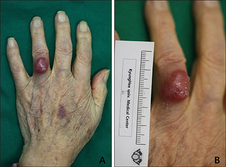
Fig. 1 (A, B) Solitary 1.5×1 cm sized erythematous nodule on the dorsal surface of the left 4th finger.
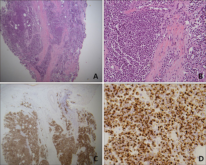
Fig. 2 Histologic findings. (A) Ill-defined tumor masses consisted of blue cells arranged in large sheets infiltrating entire dermis (H&E, ×40). (B) Uniform, atypical, small blue cells with hyperchromatic nuclei and scanty cytoplasm (H&E, ×200). (C) Immunohistochemical (IHC) staining of tumor mass in the dermis showing positive for CK-20 (IHC stain with CK-20 antibody, ×40). (D) CK-20 was stained positive in tumor cells (IHC stain with CK-20 antibody, ×400).
Reference
-
1. Hughes MP, Hardee ME, Cornelius LA, Hutchins LF, Becker JC, Gao L. Merkel cell carcinoma: epidemiology, target, and therapy. Curr Dermatol Rep. 2014; 3:46–53.
Article2. Coggshall K, Tello TL, North JP, Yu SS. Merkel cell carcinoma: an update and review: pathogenesis, diagnosis, and staging. J Am Acad Dermatol. 2018; 78:433–442.3. Chun SM, Yun SJ, Lee SC, Won YH, Lee JB. Merkel cell polyomavirus is frequently detected in Korean patients with merkel cell carcinoma. Ann Dermatol. 2013; 25:203–207.
Article4. Spalvieri C, Brunelli F, Bachmeyer CC. Merkel cell tumour of the finger. Scand J Plast Reconstr Surg Hand Surg. 2007; 41:149–151.
Article5. Ansai S, Noro S, Ogita A, Fukumoto H, Katano H, Kawana S. Case of Merkel cell carcinoma with squamous cell carcinoma possibly arising in chronic radiodermatitis of the hand. J Dermatol. 2015; 42:207–209.
Article
- Full Text Links
-
- Actions
-
Cited
- CITED
-
- Close
- Share
-
- Similar articles
-
- Merkel Cell Carcinoma
- A Case of Merkel Cell Carcinoma Concurrent with Bowen's Disease
- Primary Merkel cell carcinoma of the salivary gland: a clinicopathologic study of four cases with a review of literature
- A Case of Merkel Cell Carcinoma in the Auricle
- Cervical Spinal Metastasis of Merkel Cell Carcinoma
