Acute Phlegmonous Esophagogastritis
- Affiliations
-
- 1Department of Internal Medicine, Jeju National University School of Medicine, Jeju, Korea. sunjinboo@jejunu.ac.kr
- 2Department of Radiology, Jeju National University School of Medicine, Jeju, Korea.
- KMID: 2443639
- DOI: http://doi.org/10.4166/kjg.2019.73.4.239
Abstract
- No abstract available.
MeSH Terms
Figure
-

Fig. 1 Initial chest CT. CT scans with axial (A), sagittal (B) and coronal (C, D) images show diffuse wall thickening with intramural low density (arrows) from esophagus to gastric cardia. CT, computed tomography.
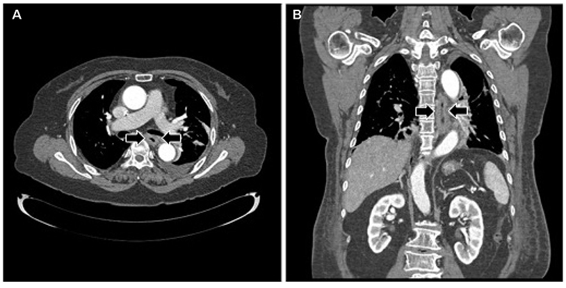
Fig. 2 Follow-up chest CT (3 weeks after medical therapy). CT scans with axial (A) and coronal (B) images reveal the improvement of diffuse wall thickening (arrows) from esophagus to gastric cardia. CT, computed tomography.

Fig. 3 Esophagographic finding (25 days after medical therapy). Esophagogram shows extraluminal barium collection (arrows) due to mucosal defects in the upper and middle esophagus.

Fig. 4 Esophagogastroduodenoscopic finding (26 days after medical therapy). Endoscopy reveals (A) a deep and round healing ulcer (white arrows) in the middle esophagus and (B) a shallow and geographic healing ulcer (black arrows) with inflammatory polyps in the upper esophagus.
Reference
-
1. Huang YC, Cheng CY, Liao CY, Hsueh C, Tyan YS, Ho SY. A rare case of acute phlegmonous esophagogastritis complicated with hypopharyngeal abscess and esophageal perforation. Am J Case Rep. 2017; 18:125–130.
Article2. Kim GY, Ward J, Henessey B, et al. Phlegmonous gastritis: case report and review. Gastrointest Endosc. 2005; 61:168–174.
Article3. Hsu CY, Liu JS, Chen DF, Shih CC. Acute diffuse phlegmonous esophagogastritis: report of a survived case. Hepatogastroenterology. 1996; 43:1347–1352.4. Jung C, Choi YW, Jeon SC, Chung WS. Acute diffuse phlegmonous esophagogastritis: radiologic diagnosis. AJR Am J Roentgenol. 2003; 180:862–863.
Article5. Kim HS, Hwang JH, Hong SS, et al. Acute diffuse phlegmonous esophagogastritis: a case report. J Korean Med Sci. 2010; 25:1532–1535.
Article6. Yun CH, Cheng SM, Sheu CI, Huang JK. Acute phlegmonous esophagitis: an unusual case (2005: 8b). Eur Radiol. 2005; 15:2380–2381.
Article7. Woo WG, Do YW, Lee GD, Lee SS. Phlegmonous esophagitis treated with internal drainage and feeding jejunostomy. Korean J Thorac Cardiovasc Surg. 2017; 50:453–455.
Article8. Hu DC, McGrath KM, Jowell PS, Killenberg PG. Phlegmonous gastritis: successful treatment with antibiotics and resolution documented by EUS. Gastrointest Endosc. 2000; 52:793–795.
Article9. Kim NY, Park JS, Lee KJ, Yun HK, Kim JS. A case of acute phlegmonous gastritis causing gastroparesis and cured with medical treatment alone. Korean J Gastroenterol. 2011; 57:309–314.
Article10. Chang PC, Wang WL, Hwang TZ, Cheng YJ. Intramural dissection with mucosal rupture alleviating phlegmonous esophagitis. Eur J Cardiothorac Surg. 2012; 41:442–444.
Article
- Full Text Links
-
- Actions
-
Cited
- CITED
-
- Close
- Share
-
- Similar articles
-
- Acute Diffuse Phlegmonous Esophagogastritis: A Case Report
- Treatment of phlegmonous esophagitis in various patients: a case series
- Two Cases of Phlegmonous Esophagogastritis in New Onset Type 2 Diabetes
- Two Cases of Acute Phlegmonous Gastritis
- Acute Phlegmonous Esophagitis with Mediastinitis Complicated by an Esophageal Perforation: A Case Report
