Dement Neurocogn Disord.
2015 Jun;14(2):94-97. 10.12779/dnd.2015.14.2.94.
Splenial Corpus Callosum Infarction Presenting with Unilateral Prosopometamorphopsia: A Case Report
- Affiliations
-
- 1Department of Neurology, Dankook University College of Medicine, Cheonan, Korea. nrdoc@dku.edu
- KMID: 2443008
- DOI: http://doi.org/10.12779/dnd.2015.14.2.94
Abstract
- BACKGROUND
Prosopometamorphopsia is a disorder of face perception in which faces appear distorted to the perceiver. Cases with unilateral prosopometamorphopsia caused by splenial lesion have been very rarely reported.
CASE REPORT
A 52-year-old right-handed woman complained that the left half of people's faces looked distorted. She stated that objects other than the face looked normal. Brain magnetic resonance imaging revealed an infarction of the left splenium of the corpus callosum. Electroencephalography and automated perimetry were normal.
CONCLUSIONS
The mechanism of unilateral prosopometamorphopsia remains unclear. However, it could be a dominant hemisphere-specific disconnection sign.
Keyword
MeSH Terms
Figure
-

Fig. 1 Illustration and drawing of facial distortions. A: The illustration was modified according to the patient's statement. B: The figure was drawn by the patient 1 year after onset.
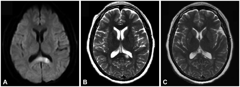
Fig. 2 Axial brain MR images of the patient. A: Diffusion-weighted image demonstrates a high signal intensity lesion in the left splenium. B: T2-weighted image obtained 1 month after onset shows a tissue loss in the left splenial lesion. C: Follow-up T2-weighted image 1 year later shows an atrophic change of the left splenial lesion.
Reference
-
1. ffytche DH, Howard RJ. The perceptual consequences of visual loss: 'positive' pathologies of vision. Brain. 1999; 122(Pt 7):1247–1260.
Article2. Bodamer J. Die prosop-agnosie. Arch Psychiatr Nervenkr Z Gesamte Neurol Psychiatr. 1947; 118:6–53.
Article3. Dalrymple KA, Davies-Thompson J, Oruc I, Handy TC, Barton JJ, Duchaine B. Spontaneous perceptual facial distortions correlate with ventral occipitotemporal activity. Neuropsychologia. 2014; 59:179–191.
Article4. Hwang JY, Ha SW, Cho EK, Han JH, Lee SH, Lee SY, et al. A Case of Prosopometamorphopsia Restricted to the Nose and Mouth with Right Medial Temporooccipital Lobe Infarction that Included the Fusiform Face Area. J Clin Neurol. 2012; 8:311–313.
Article5. Ebata S, Ogawa M, Tanaka Y, Mizuno Y, Yoshida M. Apparent reduction in the size of one side of the face associated with a small retrosplenial haemorrhage. J Neurol Neurosurg Psychiatry. 1991; 54:68–70.
Article6. Miwa H, Kondo T. Metamorphopsia restricted to the right side of the face associated with a right temporal lobe lesion. J Neurol. 2007; 254:1765–1767.
Article7. Trojano L, Conson M, Salzano S, Manzo V, Grossi D. Unilateral left prosopometamorphopsia: a neuropsychological case study. Neuropsychologia. 2009; 47:942–948.
Article8. Cho JY, Moon SY, Hong KS, Cho YJ, Kim SC, Hwang SI, et al. Teaching NeuroImages: Unilateral prosopometamorphopsia as a dominant hemisphere-specific disconnection sign. Neurology. 2011; 76:e110.
Article9. Ganssauge M, Papageorgiou E, Schiefer U. Facial dysmorphopsia: a notable variant of the "thin man" phenomenon? Graefes Arch Clin Exp Ophthalmol. 2012; 250:1491–1497.
Article10. Saito Y, Matsunaga A, Yamamura O, Ikawa M, Hamano T, Yoneda M. [A case of left hemi-facial metamorphopsia induced by infarction of the right side of the splenium of the corpus callosum]. Rinsho Shinkeigaku. 2014; 54:637–642.
Article11. Hishizawa M, Tachibana N, Hamano T. [A case of left hemifacial metamorphopsia by a right retrosplenial infarction]. Rinsho Shinkeigaku. 2015; 55:87–90.
Article12. Haxby JV, Hoffman EA, Gobbini MI. The distributed human neural system for face perception. Trends Cogn Sci. 2000; 4:223–233.
Article13. Rossion B, Caldara R, Seghier M, Schuller AM, Lazeyras F, Mayer E. A network of occipito-temporal face-sensitive areas besides the right middle fusiform gyrus is necessary for normal face processing. Brain. 2003; 126(Pt 11):2381–2395.
Article14. Gschwind M, Pourtois G, Schwartz S, Van De Ville D, Vuilleumier P. White-matter connectivity between face-responsive regions in the human brain. Cereb Cortex. 2012; 22:1564–1576.
Article15. Putnam MC, Steven MS, Doron KW, Riggall AC, Gazzaniga MS. Cortical projection topography of the human splenium: hemispheric asymmetry and individual differences. J Cogn Neurosci. 2010; 22:1662–1669.
Article16. Bukowski H, Dricot L, Hanseeuw B, Rossion B. Cerebral lateralization of face-sensitive areas in left-handers: only the FFA does not get it right. Cortex. 2013; 49:2583–2589.
Article17. Lee JI, Kim JH, Lee BH, Kim GM, Kim SH, Yoon DS, et al. Dominance specific visual extinction associated with callosal disconnection. Neurocase. 2010; 16:7–14.
Article
- Full Text Links
-
- Actions
-
Cited
- CITED
-
- Close
- Share
-
- Similar articles
-
- A Case of Transient Isolated Splenial Lesion of the Corpus Callosum After New Onset Seizure
- Transient Splenial Lesion of the Corpus Callosum in Patients with Infectious Disease
- Reversible Splenial Lesion in the Corpus Callosum on MRI after Ingestion of a Herbicide Containing Glufosinate Ammonium: A Case Report
- Lithium-Induced Downbeat Nystagmus with Reversible Splenial Lesion
- Reversible Lesion in The splenium of The Corpus Callosum Induced by Topiramate in a Patient with Migraine
