Progressive Right Ventricular Aneurysm in a Patient with Systemic Sarcoidosis
- Affiliations
-
- 1Department of Cardiovascular Medicine, Graduate School of Medicine, The University of Tokyo, Tokyo, Japan. amiyae-tky@umin.ac.jp
- KMID: 2442753
- DOI: http://doi.org/10.4250/jcvi.2019.27.e18
Abstract
- No abstract available.
MeSH Terms
Figure
-
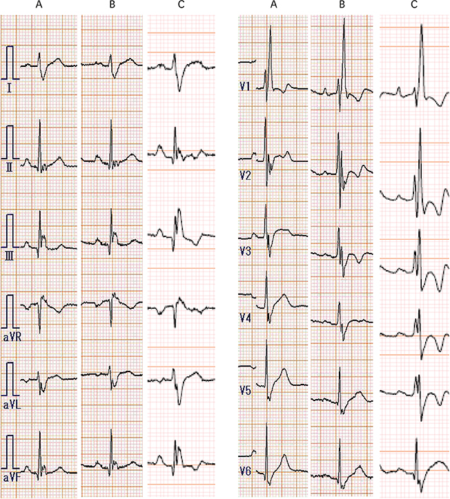
Figure 1 (A) Electrocardiogram (ECG) at the initial admission shows right bundle branch block. (B) ECG performed 8 years after the initial admission revealed a newly inverted T wave in leads V2–3. (C) At the current admission, the ECG shows a newly inverted T wave in leads II, III, aVF, V4, and V5.
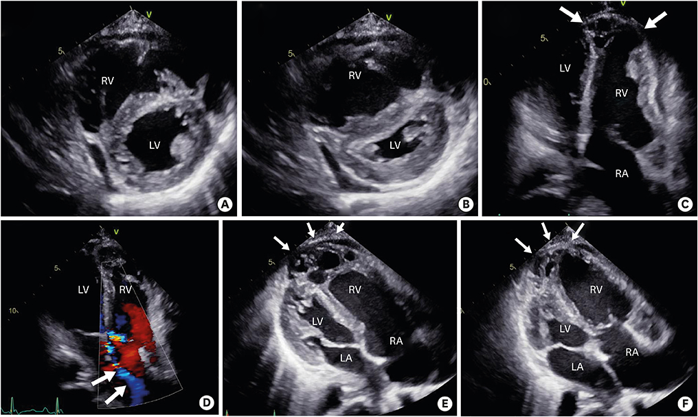
Figure 2 (A, B) A transthoracic echocardiogram in the parasternal short axis view shows severe RV dilatation and LV compression (A: diastole, B: systole). (C, D) Apical four-chamber view reveals severe RV dilatation with apical aneurysm (C: arrows) and severe tricuspid regurgitation (D: arrows). (E, F) Apical four-chamber RV-focused view also shows RV aneurysm in diastole (E: arrows) and systole (F: arrows). LA: left atrium, LV: left ventricle, RA: right atrium, RV: right ventricle.
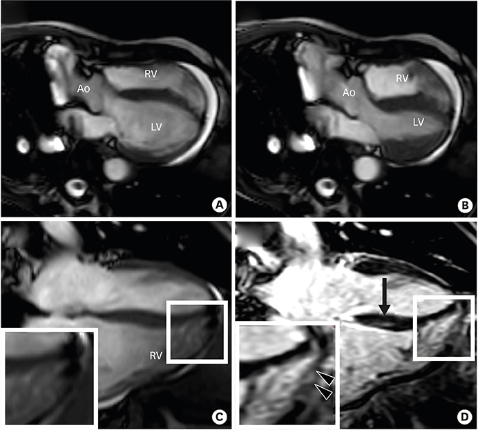
Figure 3 Functional (cine) cardiac magnetic resonance images in the four-chamber view in diastole (A) and systole (B) show normal LV function, RV dilatation and RV apical aneurysm. The corresponding cine (C) and contrast-enhanced magnetic resonance image (D) in the four-chamber view show late gadolinium enhancement in the RV apical wall (triangles) and basal septum (arrows). Ao: aorta, LV: left ventricle, PA: pulmonary artery, RV: right ventricle.
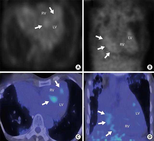
Figure 4 A [18F]fluorodeoxyglucose positron emission tomographic scan demonstrates focal enhancement of the intraventricular septum in the axial view (A and C: arrows) and in the coronal plane (B and D: arrows). LV: left ventricle, RV: right ventricle.
- Full Text Links
-
- Actions
-
Cited
- CITED
-
- Close
- Share
-
- Similar articles
-
- A Case of Ichthyosiform Sarcoidosis
- Heart Transplantation Performed in a Patient with Isolated Cardiac Sarcoidosis
- Systemic Sarcoidosis Presenting as Bilateral Optic Neuritis
- Systemic Sarcoidosis Diagnosed through Plantar Skin Lesion in a Chronic Cough Patient
- A Case of Cutaneous Sarcoidosis in the Mucosal Membrane of the Lower Lip
