Coronary Computed Tomography Angiography for the Diagnosis of Vasospastic Angina: Comparison with Invasive Coronary Angiography and Ergonovine Provocation Test
- Affiliations
-
- 1Department of Internal Medicine and Cardiovascular Center, Seoul National University Hospital, Seoul, Korea. cardiman73@gmail.com
- 2Department of Radiology, Seoul National University Hospital, Seoul, Korea. iameuna1@gmail.com
- KMID: 2442706
- DOI: http://doi.org/10.3348/kjr.2018.0847
Abstract
OBJECTIVE
To investigate the diagnostic validity of coronary computed tomography angiography (cCTA) in vasospastic angina (VA) and factors associated with discrepant results between invasive coronary angiography with the ergonovine provocation test (iCAG-EPT) and cCTA.
MATERIALS AND METHODS
Of the 1397 patients diagnosed with VA from 2006 to 2016, 33 patients (75 lesions) with available cCTA data from within 6 months before iCAG-EPT were included. The severity of spasm (% diameter stenosis [%DS]) on iCAG-EPT and cCTA was assessed, and the difference in %DS (Δ%DS) was calculated. Δ%DS was compared after classifying the lesions according to pre-cCTA-administered sublingual nitroglycerin (SL-NG) or beta-blockers. The lesions were further categorized with %DS ≥ 50% on iCAG-EPT or cCTA defined as a significant spasm, and the diagnostic performance of cCTA on identifying significant spasm relative to iCAG-EPT was assessed.
RESULTS
Compared to lesions without SL-NG treatment, those with SL-NG treatment showed a higher Δ%DS (39.2% vs. 22.1%, p = 0.002). However, there was no difference in Δ%DS with or without beta-blocker treatment (35.1% vs. 32.6%, p = 0.643). The significant difference in Δ%DS associated with SL-NG was more prominent in patients who were aged < 60 years, were male, had body mass index < 25 kg/m2, and had no history of hypertension, diabetes, or dyslipidemia. Based on iCAG-EPT as the reference, the per-lesion-based sensitivity, specificity, positive predictive value, negative predictive value, and accuracy of cCTA for VA diagnosis were 7.5%, 94.0%, 60.0%, 47.1%, and 48.0%, respectively.
CONCLUSION
For patients with clinically suspected VA, confirmation with iCAG-EPT needs to be considered without completely excluding the diagnosis of VA simply based on cCTA results, although further prospective studies are required for confirmation.
Keyword
MeSH Terms
Figure
-
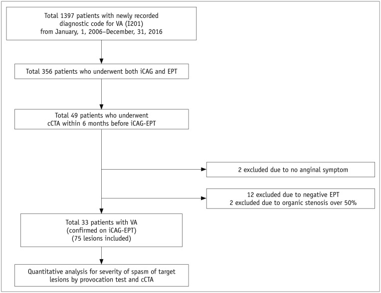
Fig. 1 Flow diagram showing study design and patient selection.cCTA = coronary computed tomography angiography, EPT = ergonovine provocation test, iCAG = invasive coronary angiography, VA = vasospastic angina
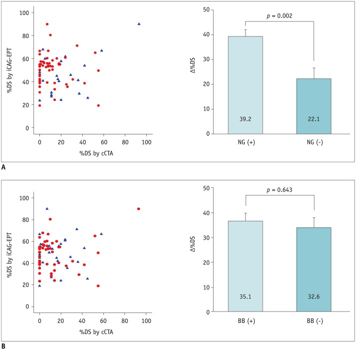
Fig. 2 Effect of pre-medication on difference in detecting severity of spasm between cCTA and iCAG-EPT.A. SL-NG. B. BBs. BB = beta-blocker, iCAG-EPT = iCAG with EPT, SL-NG = sublingual nitroglycerine, %DS = % diameter stenosis, Δ%DS = difference in %DS
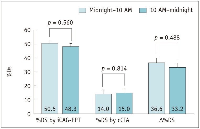
Fig. 3 Difference in severity of spasm between lesions according to time (from midnight to 10 AM or 10 AM to midnight) when cCTA was performed.
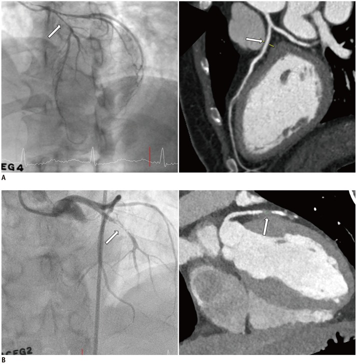
Fig. 4 Representative cases of Matched-Insignificant (A) and Matched-Significant (B) vasospasm between cCTA and iCAG-EPT.
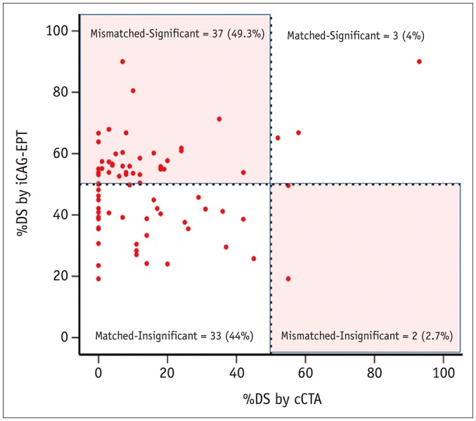
Fig. 5 Concordance or discordance between cCTA and iCAG-EPT in differentiating significant from insignificant vasospasm with use of 50% cutoff value.
Reference
-
1. JCS Joint Working Group. Guidelines for diagnosis and treatment of patients with vasospastic angina (coronary spastic angina) (JCS 2013). Circ J. 2014; 78:2779–2801. PMID: 25273915.2. Kim HL, Kim J, Kim HJ, Lim WH, Lee JY. Incidence and factors associated with mortality in 2,476 patients with variant angina in Korea. Sci Rep. 2017; 7:46031. PMID: 28383055.
Article3. Levenson B, Albrecht A, Göhring S, Haerer W, Reifart N, Ringwald G, et al. [6th report of the German Association of Cardiologists in private practice (BNK) on quality assurance in cardiac catheterization and coronary intervention 2006–2009]. Herz. 2011; 36:41–49. PMID: 21308430.4. Cho I, Al'Aref SJ, Berger A, Ó Hartaigh B, Gransar H, Valenti V, et al. Prognostic value of coronary computed tomographic angiography findings in asymptomatic individuals: a 6-year follow-up from the prospective multicentre international CONFIRM study. Eur Heart J. 2018; 39:934–941. PMID: 29365193.
Article5. Raff GL, Chinnaiyan KM, Cury RC, Garcia MT, Hecht HS, Hollander JE, et al. SCCT guidelines on the use of coronary computed tomographic angiography for patients presenting with acute chest pain to the emergency department: a report of the Society of Cardiovascular Computed Tomography Guidelines Committee. J Cardiovasc Comput Tomogr. 2014; 8:254–271. PMID: 25151918.
Article6. Ferencik M, Mayrhofer T, Bittner DO, Emami H, Puchner SB, Lu MT, et al. Use of high-risk coronary atherosclerotic plaque detection for risk stratification of patients with stable chest pain: a secondary analysis of the promise randomized clinical trial. JAMA Cardiol. 2018; 3:144–152. PMID: 29322167.7. Abbara S, Blanke P, Maroules CD, Cheezum M, Choi AD, Han BK, et al. SCCT guidelines for the performance and acquisition of coronary computed tomographic angiography: a report of the society of cardiovascular Computed Tomography Guidelines Committee: endorsed by the North American Society for Cardiovascular Imaging (NASCI). J Cardiovasc Comput Tomogr. 2016; 10:435–449. PMID: 27780758.8. Levine GN, Bates ER, Blankenship JC, Bailey SR, Bittl JA, Cercek B, et al. 2011 ACCF/AHA/SCAI guideline for percutaneous coronary intervention. A report of the American College of Cardiology Foundation/American Heart Association task force on practice guidelines and the society for cardiovascular angiography and interventions. J Am Coll Cardiol. 2011; 58:e44–e122. PMID: 22070834.9. Takagi Y, Yasuda S, Takahashi J, Tsunoda R, Ogata Y, Seki A, et al. Clinical implications of provocation tests for coronary artery spasm: safety, arrhythmic complications, and prognostic impact: multicentre registry study of the Japanese Coronary Spasm Association. Eur Heart J. 2013; 34:258–267. PMID: 22782943.
Article10. Moon JH, Park EA, Lee W, Yin YH, Chung JW, Park JH, et al. The diagnostic accuracy, image quality and radiation dose of 64-slice dual-source CT in daily practice: a single institution's experience. Korean J Radiol. 2011; 12:308–318. PMID: 21603290.
Article11. Park EA, Lee W, Kim KW, Kim KG, Thomas A, Chung JW, et al. Iterative reconstruction of dual-source coronary CT angiography: assessment of image quality and radiation dose. Int J Cardiovasc Imaging. 2012; 28:1775–1786. PMID: 22187198.
Article12. Ito T, Terashima M, Kaneda H, Nasu K, Ehara M, Kinoshita Y, et al. In vivo assessment of ergonovine-induced coronary artery spasm by 64-slice multislice computed tomography. Circ Cardiovasc Imaging. 2012; 5:226–232. PMID: 22342944.
Article13. Shin DI, Baek SH, Her SH, Han SH, Ahn Y, Park KH, et al. The 24-month prognosis of patients with positive or intermediate results in the intracoronary ergonovine provocation test. JACC Cardiovasc Interv. 2015; 8:914–923. PMID: 26003026.
Article14. Dwivedi A, Al'Aref SJ, Lin FY, Min JK. Evaluation of atherosclerotic plaque in non-invasive coronary imaging. Korean Circ J. 2018; 48:124–133. PMID: 29441745.
Article15. Dewey M, Rief M, Martus P, Kendziora B, Feger S, Dreger H, et al. Evaluation of computed tomography in patients with atypical angina or chest pain clinically referred for invasive coronary angiography: randomised controlled trial. BMJ. 2016; 355:i5441. PMID: 27777234.
Article16. Song JK, Park SW, Kang DH, Hong MK, Kim JJ, Lee CW, et al. Safety and clinical impact of ergonovine stress echocardiography for diagnosis of coronary vasospasm. J Am Coll Cardiol. 2000; 35:1850–1856. PMID: 10841234.
Article17. Kang EJ, Kim MH, De Jin C, Seo J, Kim DW, Yoon SK, et al. Noninvasive detection of coronary vasospastic angina using a double-acquisition coronary CT angiography protocol in the presence and absence of an intravenous nitrate: a pilot study. Eur Radiol. 2017; 27:1136–1147. PMID: 27380904.
Article18. Kang KM, Choi SI, Chun EJ, Kim JA, Youn TJ, Choi DJ. Coronary vasospastic angina: assessment by multidetector CT coronary angiography. Korean J Radiol. 2012; 13:27–33. PMID: 22247633.
Article19. Jin CD, Kim MH, Kang EJ, Cho YR, Park TH, Lee KN, et al. Assessing vessel tone during coronary artery spasm by dual-acquisition multidetector computed tomography angiography. Cardiology. 2018; 139:25–32. PMID: 29166637.
Article20. Ito K, Ogawa T, Yoshimura M. Severe coronary spasm occasionally detected by coronary computed tomography. Eur Heart J. 2009; 30:2768. PMID: 19710074.
Article21. Nakahara T, Toyama T, Tsushima Y, Kurabayashi M. Coronary vasospasm during CT angiography. J Cardiovasc Comput Tomogr. 2014; 8:328–330. PMID: 25065860.
Article22. Hong MK, Park SW, Lee CW, Ko JY, Kang DH, Song JK, et al. Intravascular ultrasound findings of negative arterial remodeling at sites of focal coronary spasm in patients with vasospastic angina. Am Heart J. 2000; 140:395–401. PMID: 10966536.
Article23. López-Sendón J, Swedberg K, McMurray J, Tamargo J, Maggioni AP, Dargie H, et al. Task ForceOn Beta-Blockers of the European Society of Cardiology. Expert consensus document on beta-adrenergic receptor blockers. Eur Heart J. 2004; 25:1341–1362. PMID: 15288162.24. Hadi HA, Suwaidi JA. Endothelial dysfunction in diabetes mellitus. Vasc Health Risk Manag. 2007; 3:853–876. PMID: 18200806.25. Jansson PA. Endothelial dysfunction in insulin resistance and type 2 diabetes. J Intern Med. 2007; 262:173–183. PMID: 17645585.
Article26. Kugiyama K, Ohgushi M, Motoyama T, Sugiyama S, Ogawa H, Yoshimura M, et al. Nitric oxide-mediated flow-dependent dilation is impaired in coronary arteries in patients with coronary spastic angina. J Am Coll Cardiol. 1997; 30:920–926. PMID: 9316519.
Article
- Full Text Links
-
- Actions
-
Cited
- CITED
-
- Close
- Share
-
- Similar articles
-
- Diagnostic Significance of ECG Ergonovine Provocation Test in Patients with Vasospastic Angina
- The Vasomotor Tone In Vasospastic Angina
- Exercise-Induced Vasospastic Angina With Prominent Regional Wall Motion Abnormality
- Follow-up Provocation Test in Patients with Coronary Artery Spasm
- Does a Negative Ergonovine Provocation Test Truly Predict Freedom from Variant Angina?
