Ansa Pancreatica-Type Anatomic Variation of the Pancreatic Duct in Patients with Recurrent Acute Pancreatitis and Chronic Localized Pancreatitis
- Affiliations
-
- 1Department of Radiology and Research Institute of Radiology, University of Ulsan College of Medicine, Asan Medical Center, Seoul, Korea. medimash@gmail.com
- KMID: 2442528
- DOI: http://doi.org/10.3348/jksr.2019.80.2.365
Abstract
- Ansa pancreatic is a rare variation of pancreas duct. Ansa pancreatica is characterized by focal accessory duct atrophy and an additional curved duct linking main and accessory ducts replacing atrophied duct. Ansa pancreatica is considered as a predisposing factor of recurrent pancreatitis. Pancreatitis can be localized in pancreas head and uncinate process, because pancreas head and uncinate process might be drained through the additional hooked duct of ansa pancreatica. We reports three cases of localized chronic or recurrent pancreatitis cases with underlying ansa pancreatica type anatomic variation.
MeSH Terms
Figure
-
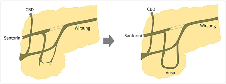
Fig. 1 Development of ansa pancreatica type anatomic variation. A curved additional duct is formed in ansa pancreatica replacing atrophied accessory duct during pancreatic duct development. The accessory duct arises from the main pancreatic duct and runs a hooked course anteriorly to the main duct ending in or around the minor papilla. CBD = common bile duct
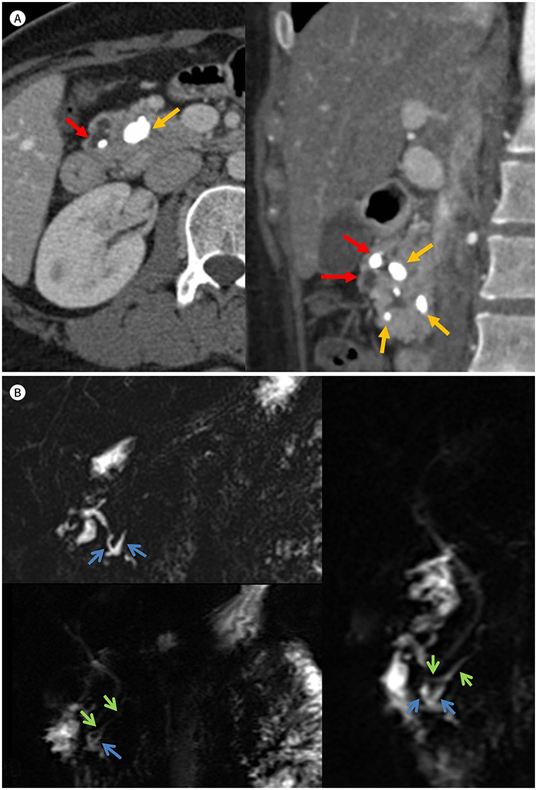
Fig. 2 A 24-year-old female patient with recurrent history of acute pancreatitis. A, B. Axial scan and coronal (A) reconstructed image of contrast-enhanced CT reveal dilated duct of Santorini with impacted pancreaticolith within the dilated duct of Santorini (red arrows). In addition, multiple pancreas parenchymal calcifications clustered in pancreatic head suggesting localized form of chronic pancreatitis (orange arrows). The coronal T2 weighted images of MRI and magnetic resonance cholangiopancreatography (B) show dilated duct of Santorini (blue arrows) with curved appearance and normal appearing duct of Wirshung (green arrows) suggesting ansa pancreatica.
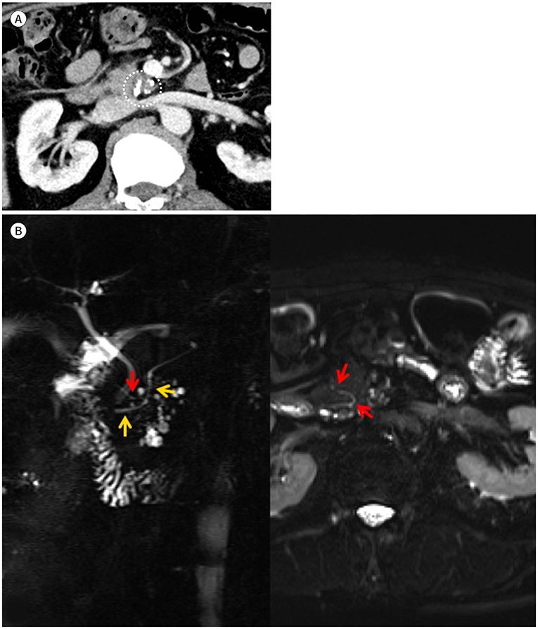
Fig. 3 A 62-year-old male patient with calcified nodule in uncinate process. A, B. The axial contrast-enhanced CT (A) reveals a small nodular lesion (circle) in the pancreas uncinate process suggesting localized chronic pancreatitis. The magnetic resonance cholangiopancreatography and axial T2 weighted images (B) reveal a hooked additional pancreatic duct arise from normal appearing main pancreatic duct (yellow arrows) draining to minor papillae (red arrows).
Reference
-
1. Prasanna LC, Rajagopal KV, Thomas HR, Bhat KM. Accessory pancreatic duct patterns and their clinical implications. J Clin Diagn Res. 2015; 9:AC05–AC07.
Article2. Dimitriou I, Katsourakis A, Nikolaidou E, Noussios G. The main anatomical variations of the pancreatic duct system: review of the literature and its importance in surgical practice. J Clin Med Res. 2018; 10:370–375.
Article3. Adibelli ZH, Adatepe M, Imamoglu C, Esen OS, Erkan N, Yildirim M. Anatomic variations of the pancreatic duct and their relevance with the Cambridge classification system: MRCP findings of 1158 consecutive patients. Radiol Oncol. 2016; 50:370–377.
Article4. Dawson W, Langman J. An anatomical-radiological study on the pancreatic duct pattern in man. Anat Rec. 1961; 139:59–68.
Article5. Bhasin DK, Rana SS, Nanda M, Gupta R, Nagi B, Wig JD. Ansa pancreatica type of ductal anatomy in a patient with idiopathic acute pancreatitis. JOP. 2006; 7:315–320.6. Kim HM, Park JY, Kim MJ. Ansa pancreatica: a case report of a type of ductal variation in a patient with idiopathic acute recurrent pancreatitis. J Korean Soc Radiol. 2010; 63:83–86.
Article7. Jarrar MS, Khenissi A, Ghrissi R, Hamila F, Letaief R. Ansa pancreatica: an anatomic variation and a rare cause of acute pancreatitis. Surg Radiol Anat. 2013; 35:745–748.
Article8. Hayashi TY, Gonoi W, Yoshikawa T, Hayashi N, Ohtomo K. Ansa pancreatica as a predisposing factor for recurrent acute pancreatitis. World J Gastroenterol. 2016; 22:8940–8948.
Article9. Borghei P, Sokhandon F, Shirkhoda A, Morgan DE. Anomalies, anatomic variants, and sources of diagnostic pitfalls in pancreatic imaging. Radiology. 2013; 266:28–36.
Article10. Ishii H, Arai K, Fukushima M, Maruoka Y, Hoshino M, Nakamura A, et al. Fusion variations of pancreatic ducts in patients with anomalous arrangement of pancreaticobiliary ductal system. J Hepatobiliary Pancreat Surg. 1998; 5:327–332.
Article
- Full Text Links
-
- Actions
-
Cited
- CITED
-
- Close
- Share
-
- Similar articles
-
- Ansa Pancreatica: A Case Report of a Type of Ductal Variation in a Patient with Idiopathic Acute Recurrent Pancreatitis
- Recurrent pancreatitis in the setting of gallbladder agenesis,ansa pancreatica, Santorinicoele and eventual intraductal papillary mucinous neoplasia (IPMN)
- Etiology, Pathogenesis and Natural Course of Chronic Pancreatitis
- A Case of Congenital Anomaly of the Pancreatic Duct Discovered in a Child with Recurrent Acute Pancreatitis
- Endoscopic Treatment of Chronic Pancreatitis
