J Korean Soc Spine Surg.
2019 Mar;26(1):21-25. 10.4184/jkss.2019.26.1.21.
Unilateral Biportal Endoscopy as a Treatment for Acute Radiculopathy after Osteoporotic Lumbar Compression Fracture: A Case Report
- Affiliations
-
- 1Department of Orthopedic Surgery, Bumin Hospital, Seoul, Korea. esshappy@daum.net
- KMID: 2442344
- DOI: http://doi.org/10.4184/jkss.2019.26.1.21
Abstract
- STUDY DESIGN: Case report.
OBJECTIVES
To document unilateral biportal endoscopy (UBE) as a treatment for acute radiculopathy after osteoporotic vertebral fracture. SUMMARY OF LITERATURE REVIEW: Acute radiculopathy after osteoporotic vertebral fracture leads to claudication. Treatment of osteoporotic vertebral fractures with accompanying radiating pain is challenging.
MATERIALS AND METHODS
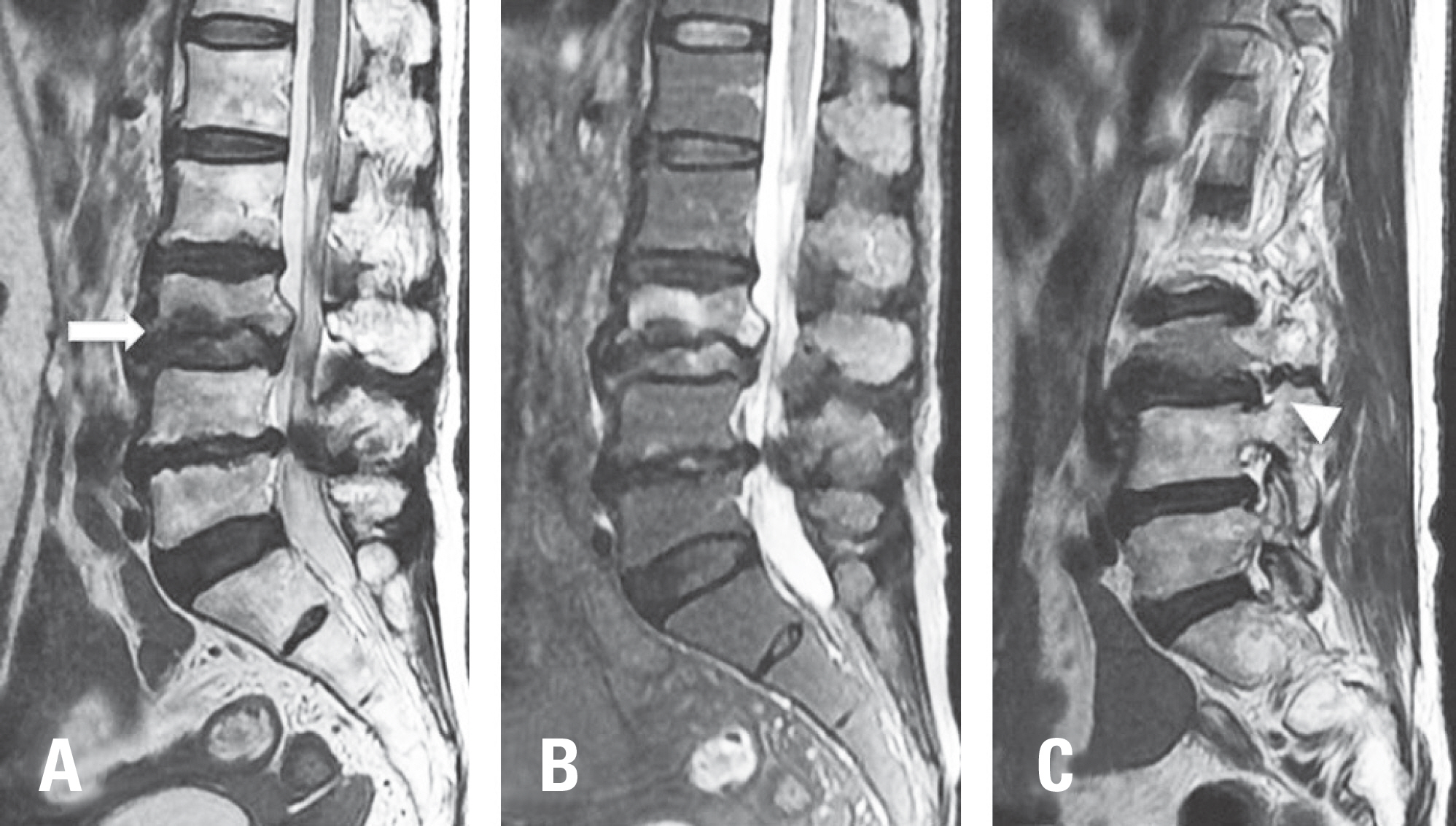
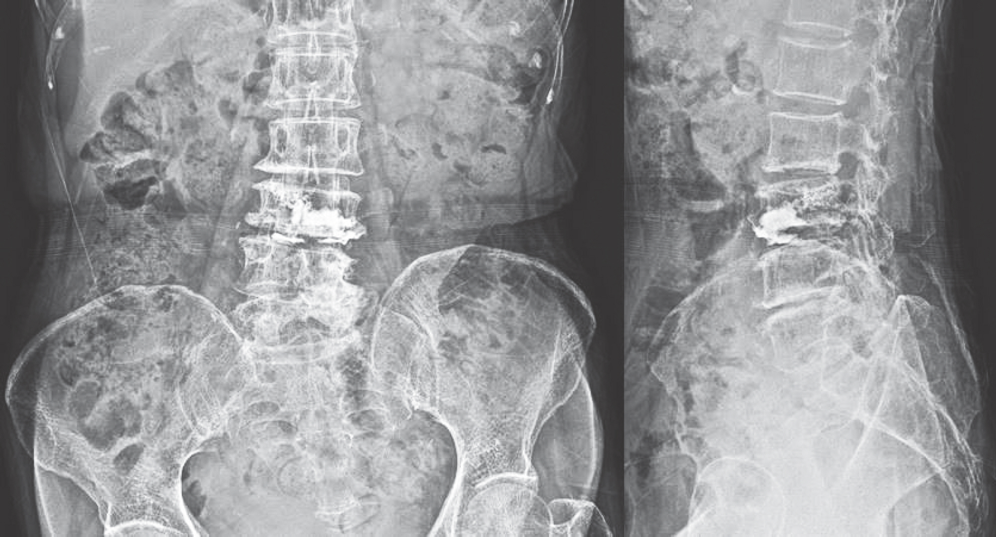
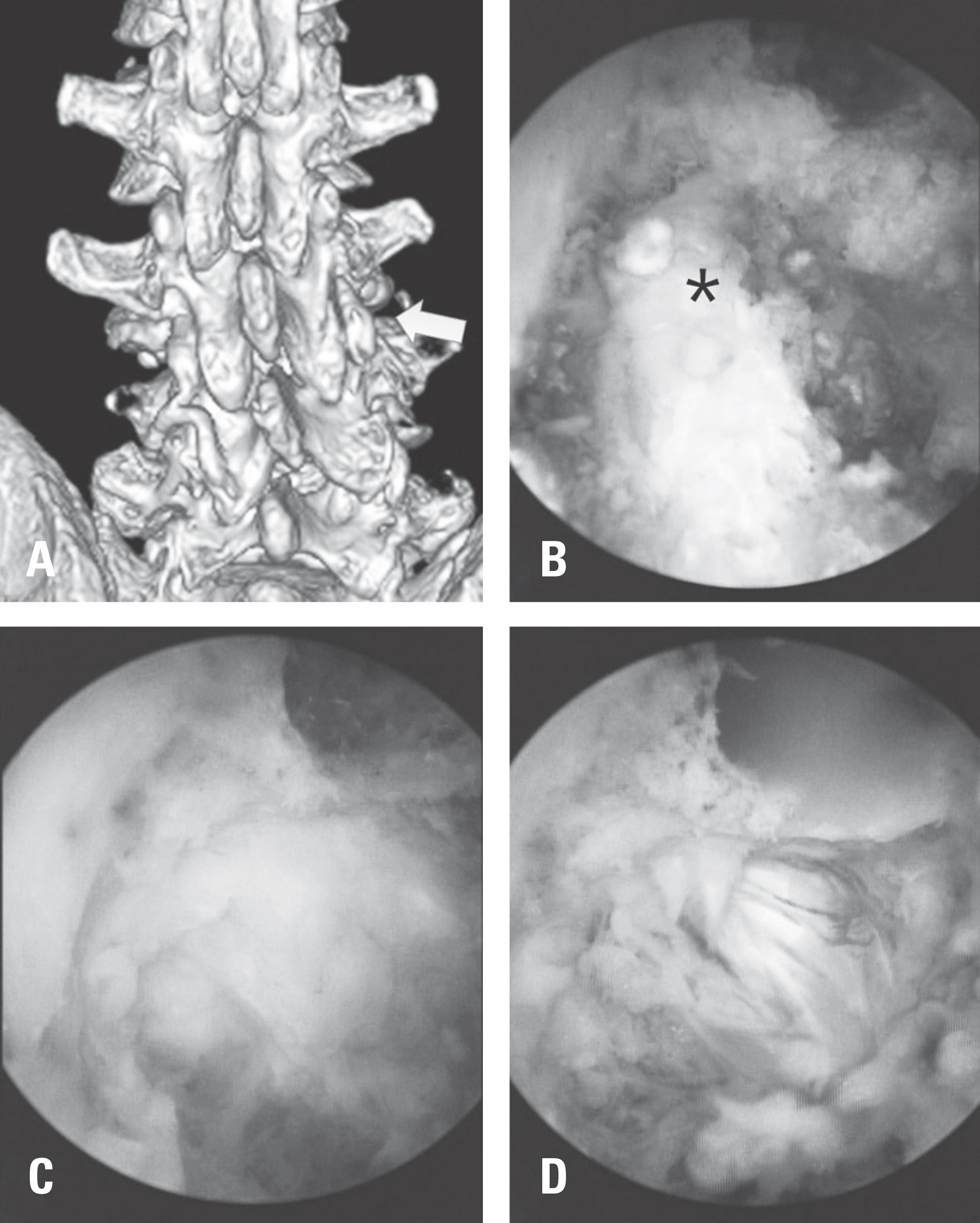
A 74-year-old woman was diagnosed with an osteoporotic vertebral fracture at L3 after slipping and falling. Vertebroplasty was performed for the osteoporotic vertebral fracture at L3. She still complained of right lower extremity radiating pain. UBE was performed to treat acute radiculopathy.
RESULTS
Foraminal decompression using UBE was performed at the L3-4 right foraminal area. Her symptoms resolved after surgery.
CONCLUSIONS
UBE is a useful treatment method for acute radiculopathy after osteoporotic vertebral fracture.
MeSH Terms
Figure
Reference
-
1. Tamayo-Orozco J, Arzac-Palumbo P, Peon-Vidales H, et al. Vertebral fractures associated with osteoporosis: patient management. Am J Med. 1997 Aug; 103(2 Suppl):44–50. DOI: 10.1016/S0002-9343 (97)90026-7.
Article2. Belkoff SM, Mathis JM, Jasper LE, et al. The biomechanics of vertebroplasty: the effect of cement volume on mechanical behavior. Spine (Phila Pa 1976). 2001 Jul; 26(14):153741.3. Buchbinder R, Osborne RH, Ebeling PR, et al. A randomized trial of vertebroplasty for painful osteoporotic vertebral fractures. N Engl J Med. 2009 Aug; 361(6):557–68. DOI: 10.1056/NEJMoa0900429.
Article4. Kim DE, Kim HS, Kim SW, et al. Clinical Analysis of Acute Radiculopathy after Osteoporotic Lumbar Compression Fracture. J Korean Neurosurg Soc. 2015 Jan; 57(1):32–5. DOI: 10.3340/jkns.2015.57.1.32.
Article5. Doo TH, Shin DA, Kim HI, et al. Clinical relevance of pain patterns in osteoporotic vertebral compression fractures. J Korean Med Sci. 2008 Dec; 23(6):1005–10. DOI: 10.3346/jkms.2008.23.6.1005.
Article6. Miller JD, Nader R. Treatment of combined osteoporotic compression fractures and spinal stenosis: use of vertebral augumentation and interspinous process spacer. Spine (Phila Pa 1976). 2008 Sep; 33(19):E717–20. DOI: 10.1097/BRS.0b013e31817f8d40.7. Eum JH, Heo DH, Son SK, et al. Percutaneous biportal endoscopic decompression for lumbar spinal stenosis: a technical note and preliminary clinical results. J Neurosurg Spine. 2016 Apr; 24(4):602–7. DOI: 10.3171/2015.7.SPINE15304.8. Choi DJ, Kim JE, Jung JT, et al. Biportal Endoscopic Spine Surgery for Various Foraminal Lesions at the Lumbosa-cral Lesion. Asian Spine J. 2018 Jun; 12(3):569–73. DOI: 10.4184/asj.2018.12.3.569.
Article
- Full Text Links
- Actions
-
Cited
- CITED
-
- Close
- Share
- Similar articles
-
- Clinical Analysis of Acute Radiculopathy after Osteoporotic Lumbar Compression Fracture
- Osteoporotic Lumbar Compression Fracture in Patient with Ankylosing Spondylitis Treated with Kyphoplasty
- Exploring Unilateral Biportal Endoscopy for Lumbar Intradural Lesions: A Technical and Video Report on Benefits and Key Considerations
- Biportal Endoscopic Lumbar Ponte Osteotomy for Kyphosis: A Technical Note
- Unilateral Biportal Endoscopic Translaminar Keyhole Approach to Treat High-grade Up-migrated Lumbar Disc Herniation: Technical Note


