Anat Cell Biol.
2019 Mar;52(1):38-42. 10.5115/acb.2019.52.1.38.
Morphological classification of the moderator band and its relationship with the anterior papillary muscle
- Affiliations
-
- 1Department of Biomedical Engineering, College of Medical Convergence, Catholic Kwandong University, Gangneung, Korea.
- 2Department of Anatomy, Catholic Kwandong University College of Medicine, Gangneung, Korea. mshur@cku.ac.kr
- KMID: 2442327
- DOI: http://doi.org/10.5115/acb.2019.52.1.38
Abstract
- This study investigated and classified the various types of moderator band (MB) in relation to the anterior papillary muscle, with the aim of providing anatomical reference information and fundamental knowledge for use when repairing the congenital defects and understanding the conduction system. The study investigated 38 formalin-fixed human hearts of both sexes obtained from donors aged 38-90 years. The MB was evident in 36 of the 38 specimens (94.7%). The morphology of the MB and its connection with the APM took various forms. The MBs that had a distinct shape were classified into three types according to their shape: cylindrical column, long and thin column, and wide and flat column. Types 2 and 3 were the most common, appearing in 15 (41.7%) and 14 (38.9%) of the 36 specimens, respectively, while type 1 was observed in seven specimens (19.4%). Type 3 was divided into subtypes based on their length. The MB usually originated from a single root (91.7%), with the remainder exhibiting double roots. The pairs of roots in the latter cases had different shapes. The originating point of the MB ranged from the supraventricular crest to the apex of the ventricle. The most-common originating point was in the middle (25 of 36 specimens, 69.4%), followed by the upper third (13.9%), the lower third (11.1%), and the top fifth (5.6%) of the interventricular septum. This study has produced fundamental anatomical and clinical information that will be useful when designing cardiac surgical procedures.
MeSH Terms
Figure
-
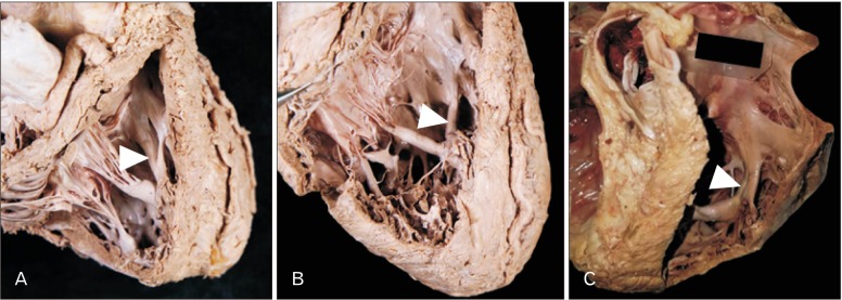
Fig. 1 Morphological classification of the moderator band (arrowheads). (A) Cylindrical column type. (B) Long and thin column type. (C) Wide and flat column type.
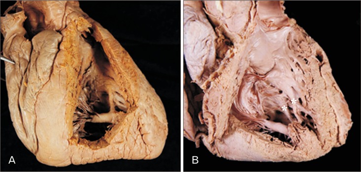
Fig. 2 Two types of roots of the moderator band (MB). (A) The MB usually originated from a single root. (B) A specimen that originated from double roots (asterisks).

Fig. 3 Branches of the moderator band (MB). The MB sometimes divided into two branches (A) or three branches (B) before attaching to the anterior wall of the right ventricle as well as the anterior papillary muscle (APM).
Reference
-
1. Kosiński A, Kozłowski D, Nowiński J, Lewicka E, Dabrowska-Kugacka A, Raczak G, Grzybiak M. Morphogenetic aspects of the septomarginal trabecula in the human heart. Arch Med Sci. 2010; 6:733–743. PMID: 22419933.2. Standring S. Gray's Anatomy: the anatomical basis of clinical practice. 41st ed. Philadelphia, PA: Elsevier Churchill Livingstone;2016.3. Loukas M, Klaassen Z, Tubbs RS, Derderian T, Paling D, Chow D, Patel S, Anderson RH. Anatomical observations of the moderator band. Clin Anat. 2010; 23:443–450. PMID: 20235167.4. Shenoy P, Lucas M, Vinay KV, Ramos A. Morphometric study of septomarginal trabeculae (moderator band). Int J Anat Res. 2016; 4:3302–3308.5. Bandeira ST, Wafae GC, Ruiz C, Nascimento SR, Fernandes JR, Wafae N. Morphological classification of the septomarginal trabecula in humans. Folia Morphol (Warsz). 2011; 70:300–304. PMID: 22117250.6. Raghavendra AY, Kavitha , Kumar A, Tarvadi P, Harsha CR. Anatomical study of the moderator band. Nitte Univ J Health Sci. 2013; 3:78–81.7. Kosiński A, Nowiński J, Kozlowski D, Piwko G, Kuta W, Grzybiak M. The crista supraventricularis in the human heart and its role in the morphogenesis of the septomarginal trabecula. Ann Anat. 2007; 189:447–456. PMID: 17910398.8. Wu MC, Ku PM, Chou MC, Huang TY. Usefulness of 64-slice computed tomography for evaluation of double-chambered right ventricle combined with atrial and ventricular septal defects. Tex Heart Inst J. 2009; 36:74–75. PMID: 19436794.9. Alva C, Ho SY, Lincoln CR, Rigby ML, Wright A, Anderson RH. The nature of the obstructive muscular bundles in double-chambered right ventricle. J Thorac Cardiovasc Surg. 1999; 117:1180–1189. PMID: 10343270.10. Wong PC, Sanders SP, Jonas RA, Colan SD, Parness IA, Geva T, Van Praagh R, Spevak PJ. Pulmonary valve-moderator band distance and association with development of double-chambered right ventricle. Am J Cardiol. 1991; 68:1681–1686. PMID: 1746472.11. Yoshimura N, Matsuhisa H, Otaka S, Kitahara J, Murakami H, Uese K, Ichida F, Misaki T. Surgical management of multiple ventricular septal defects: the role of the felt sandwich technique. J Thorac Cardiovasc Surg. 2009; 137:924–928. PMID: 19327519.12. Stellin G, Padalino M, Milanesi O, Rubino M, Casarotto D, Van Praagh R, Van Praagh S. Surgical closure of apical ventricular septal defects through a right ventricular apical infundibulotomy. Ann Thorac Surg. 2000; 69:597–601. PMID: 10735705.
- Full Text Links
-
- Actions
-
Cited
- CITED
-
- Close
- Share
-
- Similar articles
-
- Rupture of the Anterior Papillary Muscle Caused by Occlusion of the First Diagonal Branch : Report of 1 Case
- Morphological Study of the Fibularis Brevis Tendon and Plantar Aponeurosis Lateral Band Insertion on the Fifth Metatarsal Base in Korean Cadavers
- Post-traumatic tricuspid regurgitation with anterior papillary muscle rupture, corrected by papillary muscle reimplantation
- A Case of Anterior Papillary Muscle Rupture with Total Occlusion of Right Coronary Artery Resulting in Inferior Myocardial Infarction
- Papillary Fibroelastoma Originating from the Left Ventricle: A case report
