Delayed diagnosis of a primary intraosseous squamous cell carcinoma: A case report
- Affiliations
-
- 1Department of Anatomy, Biochemistry and Physiology, University of Hawai'i School of Medicine, Honolulu, HI, USA. ahmedz@hawaii.edu
- 2Department of Oral and Maxillofacial Surgery, Insurance Hospital, Suez, Egypt.
- 3Oral and Maxillofacial Medicine and Diagnostics Science, CWRU School of Dental Medicine, Cleveland, OH, USA.
- KMID: 2442112
- DOI: http://doi.org/10.5624/isd.2019.49.1.71
Abstract
- Primary intraosseous squamous cell carcinoma is a rare malignant central jaw tumor derived from odontogenic epithelial remnants. Predominantly, it affects mandible, although both jaw bones may be involved. This report describes a 60-year-old man who was initially misdiagnosed with a periapical infection related to the right lower wisdom tooth. After four months, the patient presented to a private dental clinic with a massive swelling at the right side of the mandible. Panoramic radiographs and advanced imaging revealed a lesion with complete erosion of the right ramus, which extended to the orbital floor. A biopsy from the mandibular angle revealed large pleomorphic atypical squamous cells, which is the primary microscopic feature of a poorly differentiated squamous cell carcinoma.
Keyword
MeSH Terms
Figure
-
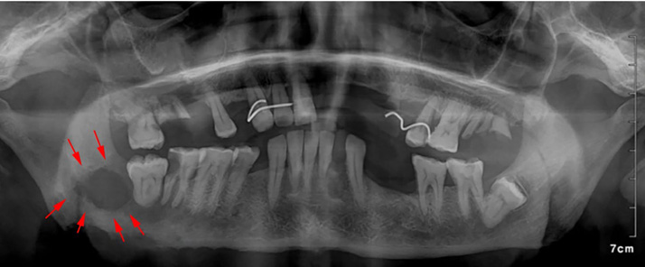
Fig. 1 Panoramic radiograph shows an ill-defined radiolucent lesion posterior to the right third molar with erosion of the inferior border of the mandible.

Fig. 2 Clinical picture over extraoral examination and panoramic radiograph. A. Supine and lateral views show a large swelling at the right side of the face. B. Panoramic radiograph shows complete erosion of the right ramus, condyle, and a portion of the mandibular body.

Fig. 3 Multi-slice computed tomography. A 3-dimensional reconstruction image shows complete erosion of the ramus. Axial, coronal, and sagittal slices show extension of the lesion (red arrows).
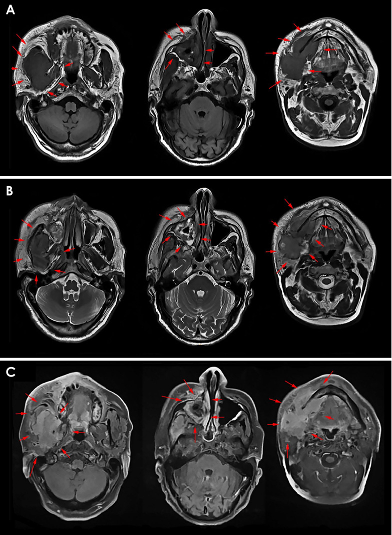
Fig. 4 Magnetic resonance imaging examination. Series of axial T1 (A), T2 (B), and post-contrast axial T1 (C) images showing the extension of the lesion (arrows) in the pterygoid region, maxillary sinus, and submandibular area (columns from left to right).

Fig. 5 Gross biopsy and histopathology examination. A. Two specimens from the angle of the mandible (3 pieces, left) and the maxillary sinus (multiple pieces, right). B. Section shows groups of large pleomorphic atypical squamous cells (arrows) with a high nucleoplasm ratio and focal intracytoplasmic keratin formation (H&E stain, ×400). C. Numerous irregularly shaped solid epithelial islands of varying sizes in the connective tissue (H&E stain, ×40). D. The basal cells are arranged in a plexiform pattern with palisading of the peripheral cells (H&E stain, ×100) (red arrows).
Reference
-
1. Bodner L, Manor E, Shear M, van der Waal I. Primary intraosseous squamous cell carcinoma arising in an odontogenic cyst: a clinicopathologic analysis of 116 reported cases. J Oral Pathol Med. 2011; 40:733–738.2. Williams MD. Update from the 4th Edition of the World Health Organization Classification of Head and Neck Tumours: Mucosal Melanomas. Head Neck Pathol. 2017; 11:110–117.
Article3. Matsuzaki H, Katase N, Matsumura T, Hara M, Yanagi Y, Nagatsuka H, et al. Solid-type primary intraosseous squamous cell carcinoma of the mandible: a case report with histopathological and imaging features. Oral Surg Oral Med Oral Pathol Oral Radiol. 2012; 114:e71–e77.
Article4. Suei Y, Tanimoto K, Taguchi A, Wada T. Primary intraosseous carcinoma: review of the literature and diagnostic criteria. J Oral Maxillofac Surg. 1994; 52:580–583.
Article5. Woolgar JA, Triantafyllou A, Ferlito A, Devaney KO, Lewis JS Jr, Rinaldo A, et al. Intraosseous carcinoma of the jaws: a clinicopathologic review. Part II: odontogenic carcinomas. Head Neck. 2013; 35:902–905.
Article6. Sengupta S, Vij H, Vij R. Primary intraosseous carcinoma of the mandible: a report of two cases. J Oral Maxillofac Pathol. 2010; 14:69–72.
Article7. Thakur A, Tupkari JV, Joy T, Gogri AA. Primary intraosseous squamous cell carcinoma—a rare odontogenic malignancy. J Oral Maxillofac Pathol. 2017; 21:320.8. Tawfik MA, Zyada MM. Odontogenic tumors in Dakahlia, Egypt: analysis of 82 cases. Oral Surg Oral Med Oral Pathol Oral Radiol Endod. 2010; 109:e67–e73.
Article9. Huang JW, Luo HY, Li Q, Li TJ. Primary intraosseous squamous cell carcinoma of the jaws. Clinicopathologic presentation and prognostic factors. Arch Pathol Lab Med. 2009; 133:1834–1840.10. Thomas G, Pandey M, Mathew A, Abraham EK, Francis A, Somanathan T, et al. Primary intraosseous carcinoma of the jaw: pooled analysis of world literature and report of two new cases. Int J Oral Maxillofac Surg. 2001; 30:349–355.
Article11. Suei Y, Taguchi A, Tanimoto K. Recommendation of modified classification for odontogenic carcinomas. Med Hypotheses. 2004; 62:382–386.
Article12. Elzay RP. Primary intraosseous carcinoma of the jaws. Review and update of odontogenic carcinomas. Oral Surg Oral Med Oral Pathol. 1982; 54:299–303.13. Reichart P, Philipsen HP. Odontogenic tumors and allied lesions. London: Quintessence Pub;2004. p. 205–225.14. Dai YL, King AD. State of the art MRI in head and neck cancer. Clin Radiol. 2018; 73:45–59.
Article15. Grisar K, Schol M, Hauben E, Schoenaers J, Politis C. Primary intraosseous squamous cell carcinoma of the mandible arising from an infected odontogenic cyst: a case report and review of the literature. Oncol Lett. 2016; 12:5327–5331.
Article16. Slootweg PJ, Müller H. Malignant ameloblastoma or ameloblastic carcinoma. Oral Surg Oral Med Oral Pathol. 1984; 57:168–176.
Article17. Waldron CA, Mustoe TA. Primary intraosseous carcinoma of the mandible with probable origin in an odontogenic cyst. Oral Surg Oral Med Oral Pathol. 1989; 67:716–724.
Article
- Full Text Links
-
- Actions
-
Cited
- CITED
-
- Close
- Share
-
- Similar articles
-
- Squamous Cell Carcinoma of the Maxilla Originated in Odontogenic Cyst: A Case Report
- Primary intraosseous carcinoma(PIOC) on mandible: Case Report
- Primary intraosseous carcinoma on mandible: A case report
- A Case of Primary Squamous Cell Carcinoma of Sigmoid Colon
- Squamous Cell Carcinoma in Bladder Diverticulum: A Report of One Case
