Fourth molar: A retrospective study and literature review of a rare clinical entity
- Affiliations
-
- 1Department of Oral and Maxillofacial Radiology, Okayama University Graduate School of Medicine, Dentistry and Pharmaceutical Sciences, Okayama, Japan. drtundebamgbose@yahoo.com
- 2Department of Oral Diagnostic Sciences, Faculty of Dentistry, Bayero University Kano, Kano, Nigeria.
- 3Department of Oral Diagnosis and Dentomaxillofacial Radiology, Okayama University Hospital, Okayama, Japan.
- 4Department of Dental Informatics, Okayama University Graduate School of Medicine, Dentistry and Pharmaceutical Sciences, Okayama, Japan.
- 5Oral Inspection and Diagnostic Center, Okayama University Hospital, Okayama, Japan.
- 6Department of Chemistry and Forensic Sciences, Savannah State University, Georgia, USA.
- KMID: 2442106
- DOI: http://doi.org/10.5624/isd.2019.49.1.27
Abstract
- PURPOSE
The prevalence of supernumerary teeth has been reported to be between 0.1% and 3.8%. The aim of this study was to determine the prevalence, clinical significance, and associated pathologies of fourth molars based on a retrospective study and a literature review.
MATERIALS AND METHODS
A 5-year retrospective prevalence study was conducted at the Department of Oral Diagnosis and Dentomaxillofacial Radiology of Okayama University Hospital, Okayama, Japan. The study involved extracting data from the digital records of patients from January 1, 2013 through December 31, 2017. The sampling frame included all patients who had panoramic radiographs, cone-beam computed tomography (CT), and multislice CT images during the period under review.
RESULTS
A total of 26,721 cases were reviewed and 87 fourth molars were identified. The prevalence of fourth molars in the 5-year study at Okayama was calculated as 0.32%. The mean age of patients with a fourth molar was 30.43 years, and the male-to-female ratio was 1:0.98. The vast majority of cases were in the maxilla (92%) and had normal shapes (89.7%); furthermore, 82.8% of cases were unerupted.
CONCLUSION
The prevalence of fourth molars in the study population was found to be 0.32%, and fourth molars occurred with approximately equal frequency in males and females. Fourth molars were more common in the maxilla and were predominantly unerupted and small.
Keyword
MeSH Terms
Figure
-
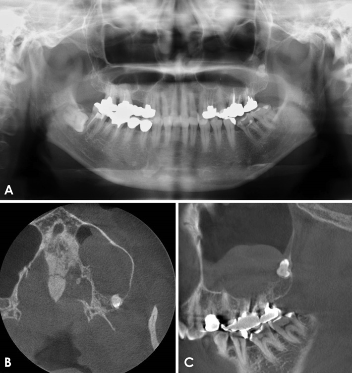
Fig. 1 A. Panoramic radiograph shows a borderline case classified as “cannot decide,” portraying a toothlike radiopacity in posterior maxillary left region that cannot be classified as a third or fourth molar because of the absence of both maxillary third molars. B and C. Conebeam computed tomographic images demonstrate an abnormally shaped tooth-like entity with 2 distinct crowns and fused roots with an associated mucus retention cyst in the posterior wall of the maxillary sinus.
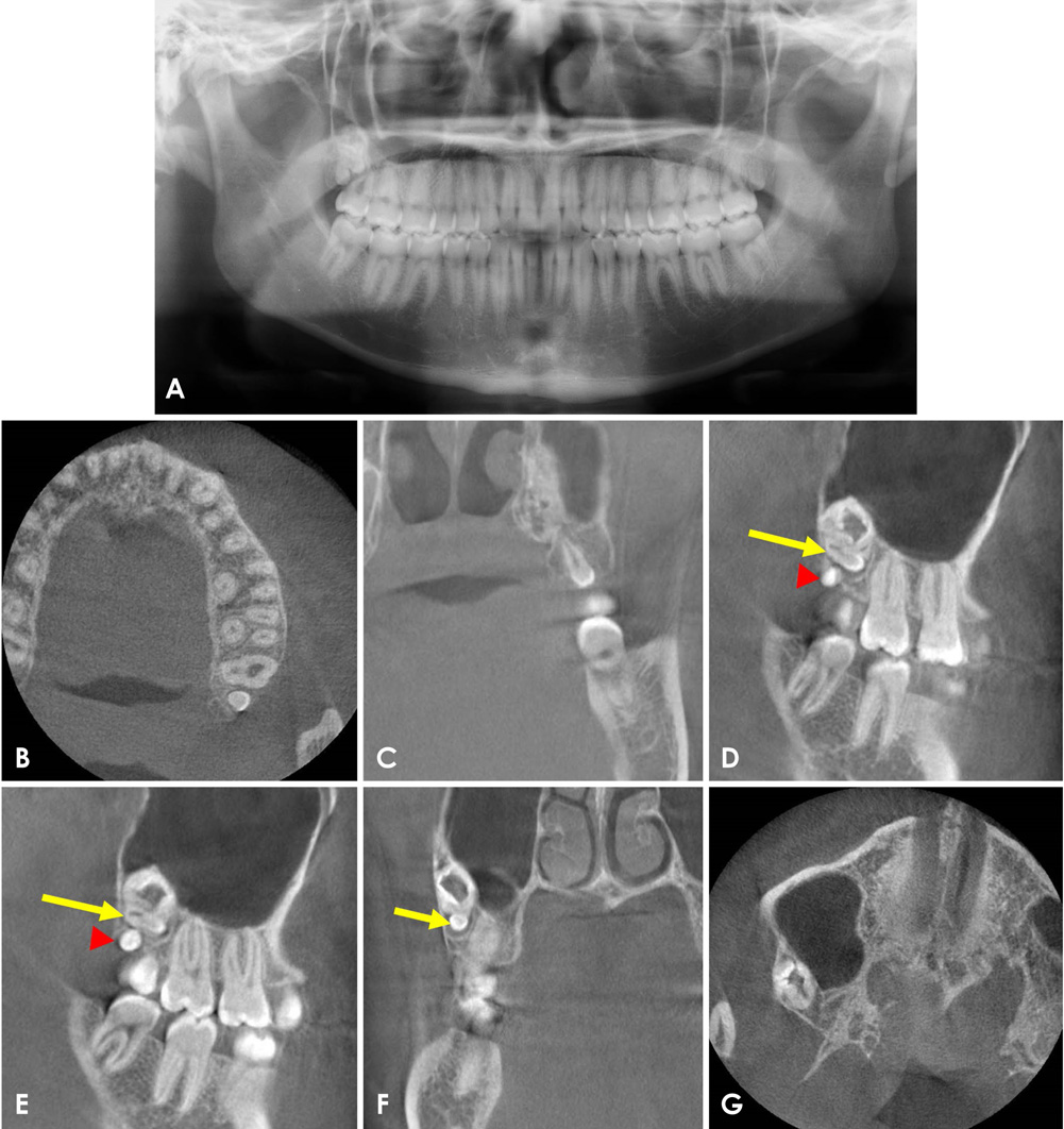
Fig. 2 A. Panoramic radiograph shows a fourth molar distal to the maxillary left third molar. There is also a fourth molar associated with a suspected odontoma in the region of the maxillary right third molar. Both fourth molars are in the maxillary tuberosity. B and C. Conebeam computed tomographic (CBCT) images demonstrate the maxillary left fourth molar as a normal-shaped microdont. D and E. Sagittal CBCT images show a complex odontoma superior to the maxillary right fourth molar (arrow head). D–G. The complex odontoma consists of a horizontally impacted microdont at its inferior segment (arrow), and the upper segment consists of various component tissues of the teeth.
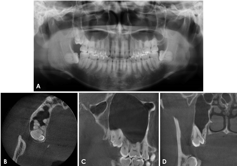
Fig. 3 A. Panoramic radiograph shows a fourth molar overlapping the maxillary right third molar. Cone-beam computed tomographic images demonstrate a fourth molar distopalatal to the maxillary right third molar (B) and a fused tooth connecting with the third molar by the dental pulp (C and D).
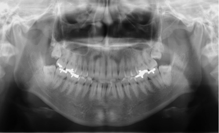
Fig. 4 Fully-formed fourth molar associated with the distal surface of the maxillary right third molar in a patient with cleidocranial dysostosis.
Reference
-
1. Pippi R. Odontomas and supernumerary teeth: is there a common origin? Int J Med Sci. 2014; 11:1282–1297.
Article2. Rajab LD, Hamdan MA. Supernumerary teeth: review of the literature and a survery of 152 cases. Int J Paediatr Dent. 2002; 12:244–254.3. Patchett CL, Crawford PJ, Cameron AC, Stephens CD. The management of supernumerary teeth in childhood - a retrospective study of practice in Bristol Dental Hospital, England and Western Dental Hospital, Sydney, Australia. Int J Paediatr Dent. 2001; 11:259–265.4. Fernández Montenegro P, Valmaseda Castellón E, Berini Aytés L, Gay Escoda C. Retrospective study of 145 supernumerary teeth. Med Oral Patol Oral Cir Bucal. 2006; 11:E339–E344.5. Harel-Raviv M, Eckler M, Raviv E, Gornitsky M. Fourth molars: a clinical study. Dent Update. 1996; 23:379–382.6. Kaya E, Güngör K, Demirel O, Özütürk Ö. Prevalence and characteristics of non-syndromic distomolars: a retrospective study. J Investig Clin Dent. 2015; 6:282–286.
Article7. Dang HQ, Constantine S, Anderson PJ. The prevalence of dental anomalies in an Australian population. Aust Dent J. 2017; 62:161–164.
Article8. Scarfe WC, Farman AG, Sukovic P. Clinical applications of cone-beam computed tomography in dental practice. J Can Dent Assoc. 2006; 72:75–80.9. Shahzad KM, Roth LE. Prevalence and management of fourth molars: a retrospective study and literature review. J Oral Maxillofac Surg. 2012; 70:272–275.
Article10. Rahnama M, Szyszkowska A, Pulawska M, Szczerba-Gwordz J. A rare case of retained fourth molar teeth in maxilla and mandible. Case report. Curr Issues Pharm Med Sci. 2014; 27:118–120.
Article11. Grimanis GA, Kyriakides AT, Spyropoulos ND. A survey on supernumerary molars. Quintessence Int. 1991; 2:989–995.12. Ruprecht A, Batniji S, el-Neweihi E. Incidence of supernumerary teeth. Ann Dent. 1984; 43:18–21.13. McMenamin JP, Hart D. Paramolars: a case report. N Y J Dent. 1985; 55:209.14. Fisher SE. Maxillary sixth molars. Br Dent J. 1982; 152:356.
Article15. Piattelli A, Tetè S. Bilateral maxillary and mandibular fourth molars. Report of a case. Acta Stomatol Belg. 1992; 89:57–60.16. Harris EF, Clark LL. An epidemiological study of hyperdontia in American blacks and whites. Angle Orthod. 2008; 78:460–465.
Article17. Batra P, Duggal R, Parkash H. Non-syndromic multiple supernumerary teeth transmitted as an autosomal dominant trait. J Oral Pathol Med. 2005; 34:621–625.
Article18. Dubuk AN, Selvig KA, Tellefsen G, Wikesjö UM. Atypically located paramolar. Report of a rare case. Eur J Oral Sci. 1996; 104:138–140.
Article
