Acute Crit Care.
2018 Nov;33(4):280-281. 10.4266/acc.2017.00598.
Tension Pneumothorax after Attempting Insertion of a Central Venous Catheter
- Affiliations
-
- 1Department of Surgery, Inje University Haeundae Paik Hospital, Inje University College of Medicine, Busan, Korea. medhun@hanmail.net
- KMID: 2441248
- DOI: http://doi.org/10.4266/acc.2017.00598
Abstract
- No abstract available.
MeSH Terms
Figure
-
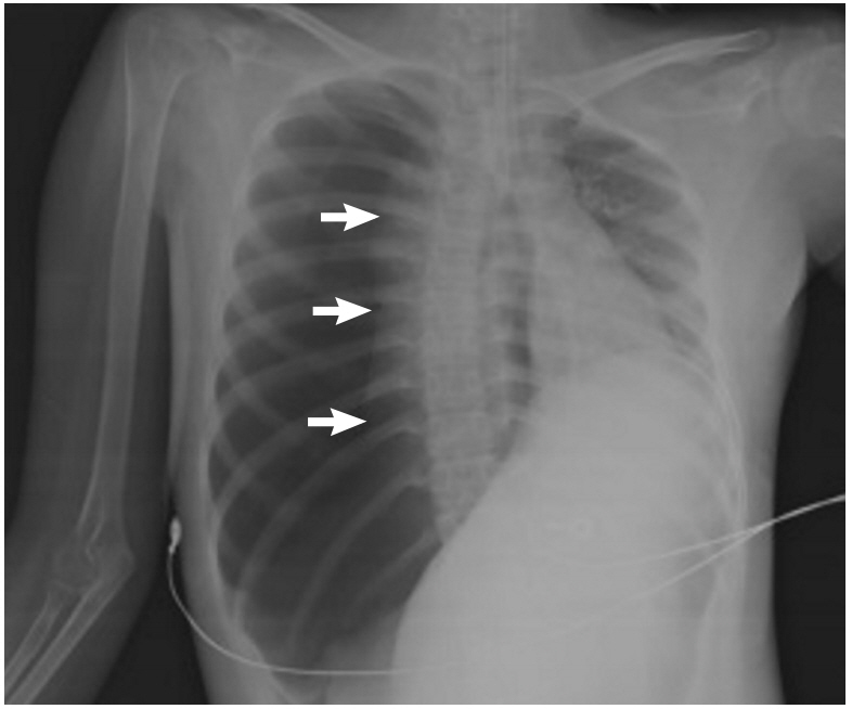
Figure 1. Chest X-ray showing right tension pneumothorax with mediastinal shifting to the left side. The right lung is completely collapsed (arrows), and the trachea is pushed to the left. The right hemidiaphragm is depressed.

Figure 2. Chest X-ray showing chest tube positioning and improved tension pneumothorax.
Reference
-
1. McGee DC, Gould MK. Preventing complications of central venous catheterization. N Engl J Med. 2003; 348:1123–33.
Article2. Eisen LA, Narasimhan M, Berger JS, Mayo PH, Rosen MJ, Schneider RF. Mechanical complications of central venous catheters. J Intensive Care Med. 2006; 21:40–6.
Article3. Roberts DJ, Leigh-Smith S, Faris PD, Blackmore C, Ball CG, Robertson HL, et al. Clinical presentation of patients with tension pneumothorax: a systematic review. Ann Surg. 2015; 261:1068–78.4. Plewa MC, Ledrick D, Sferra JJ. Delayed tension pneumothorax complicating central venous catheterization and positive pressure ventilation. Am J Emerg Med. 1995; 13:532–5.
- Full Text Links
-
- Actions
-
Cited
- CITED
-
- Close
- Share
-
- Similar articles
-
- Tension hydrothorax induced by malposition of central venous catheter: A case report
- Delayed Tension Pneumothorax Complicating Subclavian Vein Catheterization and Positive Pressure Ventilation: A case report
- Accidental Insertion of Entire Guide - Wire in the Inferior Vena Cava During Central Venous Catheterization
- Hemomediastinum Caused by Central Venous Catheter
- Delayed Tension Pneumothorax Detected 4 Days after Central Venous Catheterization: A case report
