Could Ultrasound-Guided Stimulation of Sural Nerve Affect Nerve Conduction Study?
- Affiliations
-
- 1Department of Physical Medicine and Rehabilitation, Korea University Guro Hospital, Seoul, Korea. rehab46@korea.ac.kr
- 2Radio Technology Research Department, Electronics and Telecommunications Research Institute, Daejeon, Korea.
- KMID: 2440954
- DOI: http://doi.org/10.5535/arm.2019.43.1.74
Abstract
OBJECTIVE
To determine anatomical variation of the sural nerve (SN) by ultrasonography (US) and compare sensory nerve action potential (SNAP) of the SN obtained by a control method to that obtained with adjusted method using US.
METHODS
Eighty legs of 40 healthy volunteers were enrolled. The location and formation of SN were investigated through US. Two methods of nerve conduction study (NCS) were then performed. In the control method, the cathode was placed 14 cm proximal to the lateral malleolus and the greatest SNAP amplitude was obtained by moving the cathode medially or laterally from just lateral to the calf-mid line. In adjusted NCS, the exact SN union site was stimulated in type 1. In other SN types, the stimulation was done directly over the nerve and the distance from the lateral malleolus was set to be 14 cm.
RESULTS
It was found that 73.8% of the SNs were type 1, 22.5% were direct continuation of MSCN (type 2), and 3.8% were MSCN and LSCN without communicating (type 4). However, type 3 was not found. The union point in type 1 SN was 12.6±2.5 cm proximal to the lateral malleolus and 1.4±0.7 cm lateral to the calf-midline. After stimulation adjustment, SNAP amplitude in type 1 SN was significantly increased (20.7±5.5 μV vs. 27.1±6.7 μV).
CONCLUSION
Anatomical variation of SN and its location were verified by US. US provides additional information for conducting sural NCS and helps obtain more accurate results.
Keyword
MeSH Terms
Figure
-
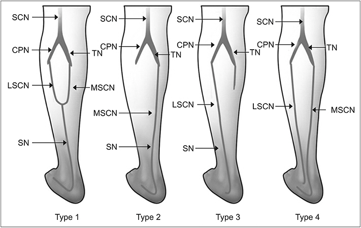
Fig. 1. Types of sural nerve formation: type 1, classical pattern (union of MSCN and LSCN); type 2, extending from MSCN alone; type 3, extending from LSCN alone; type 4, MSCN and LSCN are separate and running parallel to each other down to the ankle. SCN, sciatic nerve; CPN, common peroneal nerve; TN, tibial nerve; MSCN, medial sural cutaneous nerve; LSCN, lateral sural cutaneous nerve; SN, sural nerve.
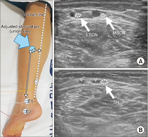
Fig. 2. Sural nerve conduction study with adjusted stimulation location and ultrasonography of a transverse view of sural nerve. (A) Traced upwardly from B level where MSCN and LSCN are just about to divide. (B) Level of ankle. MSCN, medial sural cutaneous nerve; LSCN, lateral sural cutaneous nerve; V, small saphenous vein; E1, active electrode; E2, reference electrode.
Reference
-
1. Moore KL, Dalley AF. Clinically oriented anatomy. 4th ed. Philadelphia: Lippincott Williams & Wilkins;1999.2. McMinn RM. Last’s anatomy: regional and applied. 8th ed. Edinburgh: Churchill Livingstone;1990.3. Schuchmann JA. Sural nerve conduction: a standardized technique. Arch Phys Med Rehabil. 1977; 58:166–8.4. Buschbacher RM. Sural and saphenous 14-cm antidromic sensory nerve conduction studies. Am J Phys Med Rehabil. 2003; 82:421–6.
Article5. Pyun SB, Kwon HK. The effect of anatomical variation of the sural nerve on nerve conduction studies. Am J Phys Med Rehabil. 2008; 87:438–42.
Article6. Scheidegger O, Kuffer AF, Kamm CP, Rosler KM. Reproducibility of sensory nerve conduction studies of the sural nerve using ultrasound-guided needle positioning. Muscle Nerve. 2011; 44:873–6.
Article7. Kim KH, Yoo JY, You BC. Ultrasonographic evaluation of sural nerve for nerve conduction study. Ann Rehabil Med. 2014; 38:46–51.
Article8. Mahakkanukrauh P, Chomsung R. Anatomical variations of the sural nerve. Clin Anat. 2002; 15:263–6.
Article9. Vuksanovic-Bozaric A, Radunovic M, Radojevic N, Abramovic M. The bilateral anatomical variation of the sural nerve and a review of relevant literature. Anat Sci Int. 2014; 89:57–61.
Article10. Popieluszko P, Mizia E, Henry BM, Pekala PA, Sanna B, Roy J, et al. The surgical anatomy of the sural nerve: an ultrasound study. Clin Anat. 2018; 31:450–5.
Article11. Shankar N, Selvam RP, Dhanpal N, Reddy R, Alapati A. Anatomical variations of the sural nerve in the leg: a fetal study. Neurol India. 2010; 58:24–8.
Article12. Coert JH, Dellon AL. Clinical implications of the surgical anatomy of the sural nerve. Plast Reconstr Surg. 1994; 94:850–5.
Article13. Sreenivasan A, Mansukhani KA, Sharma A, Balakrishnan L. Sural sensory nerve action potential: A study in healthy Indian subjects. Ann Indian Acad Neurol. 2016; 19:312–7.
Article14. Scheidegger O, Kihm C, Kamm CP, Rosler KM. Sural nerve conduction studies using ultrasound-guided needle positioning: influence of age and recording location. Muscle Nerve. 2016; 54:879–82.
Article15. Kimura J, Sakimura Y, Machida M, Fuchigami Y, Ishida T, Claus D, et al. Effect of desynchronized inputs on compound sensory and muscle action potentials. Muscle Nerve. 1988; 11:694–702.
Article
- Full Text Links
-
- Actions
-
Cited
- CITED
-
- Close
- Share
-
- Similar articles
-
- A Proximal Conducting Technique of Sural Nerve
- Amplitude Comparison between Sural and Distal Sural Nerves in Diabetic Neuropathy
- Determination of an Ideal Stimulation Site of the Medial Antebrachial Cutaneous Nerve Using Ultrasound and Investigation of the Efficiency
- Ultrasonographic Evaluation of Sural Nerve for Nerve Conduction Study
- A Study of Nerve Conduction Velocity of Normal Adults
