Surgical Repair of a Full-thickness Macular Hole in Retinitis Pigmentosa: a Case Report
- Affiliations
-
- 1Department of Ophthalmology, Asan Medical Center, University of Ulsan College of Medicine, Seoul, Korea. junekim@amc.seoul.kr
- KMID: 2440458
- DOI: http://doi.org/10.3341/jkos.2019.60.3.287
Abstract
- PURPOSE
To report the long-term outcome after surgical repair of a full-thickness macular hole (FTMH) in a patient with retinitis pigmentosa (RP).
CASE SUMMARY
A 55-year-old male who had been diagnosed with retinitis pigmentosa in both eyes 5 years earlier presented with decreased visual acuity in his left eye over the last 6 months. On examination, his Snellen best-corrected visual acuity (BCVA) was 1.0 in the right eye and 0.3 in the left eye. Slit-lamp examination of the anterior segment was remarkable only for posterior chamber intraocular lenses in each eye. Fundus examination demonstrated extensive bony spicule-like pigmentation in the mid-peripheral region in both eyes and a FTMH with approximately one-third disc diameter in the left eye. The optical coherence tomography (OCT) findings confirmed a FTMH with a surrounding cuff of intraretinal fluid and vitreomacular traction in the left eye. The patient underwent 23-gauge pars plana vitrectomy (PPV) with indocyanine green-assisted internal limiting membrane peeling and gas tamponade. One week postoperatively, an anatomically well-sealed macular hole was confirmed by OCT. At the 3-month postoperative follow-up, the BCVA improved to 0.63 and the hole remained closed until his last follow-up (postoperative 6 years).
CONCLUSIONS
Although macular hole is a rare occurrence in RP patients, it should be considered as a cause of significant visual loss in patients with this disorder. Our case suggested that over the long-term, PPV may be tolerable in the management for FTMH in RP.
MeSH Terms
Figure
-

Figure 1. Preoperative fundus photography images of the right (A) and left (B) eyes of the study patient are presented. Note an extensive bony spicule-like pigmentation in the midperipheral region in both eyes and a full-thickness macular hole with approximately one-third disc diameter in the left eye.
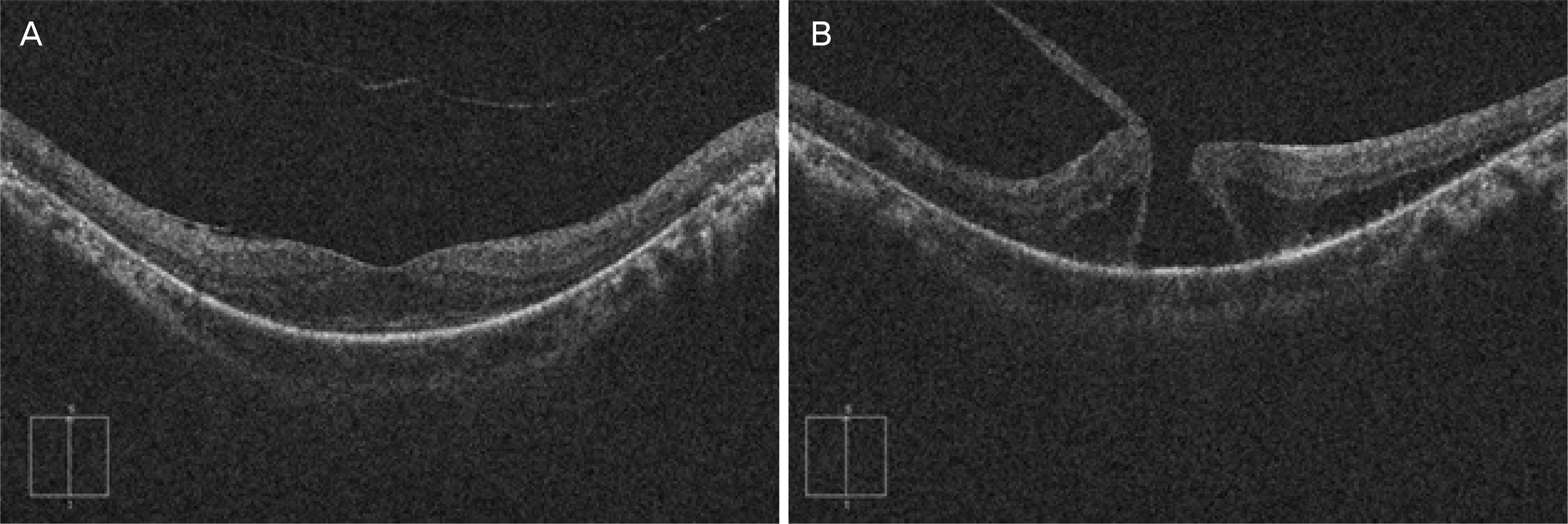
Figure 2. The preoperative optical coherence tomography images of the right (A) and left (B) eyes of the study patient are presented. Note the full-thickness macular hole with surrounding cuff of intraretinal fluid and vitreomacular traction in left eye.

Figure 3. The postoperative fundus photography and optical coherence tomography images of the left eye at 1 month (A, B), 6 years (C, D), respectively. Note that the full-thickness macular hole has resolved in each image.
Reference
-
References
1. Hartong DT, Berson EL, Dryja TP. Retinitis pigmentosa. Lancet. 2006; 368:1795–809.
Article2. You QS, Xu L, Wang YX, et al. Prevalence of retinitis pigmentosa in North China: the Beijing Eye Public Health Care Project. Acta Ophthalmol. 2013; 91:e499–500.
Article3. Hirakawa H, Iijima H, Gohdo T, Tsukahara S. Optical coherence tomography of cystoid macular edema associated with retinitis pigmentosa. Am J Ophthalmol. 1999; 128:185–91.
Article4. Testa F, Rossi S, Colucci R, et al. Macular abnormalities in Italian patients with retinitis pigmentosa. Br J Ophthalmol. 2014; 98:946–50.
Article5. Hagiwara A, Yamamoto S, Ogata K, et al. Macular abnormalities in patients with retinitis pigmentosa: prevalence on OCT examination and outcomes of vitreoretinal surgery. Acta Ophthalmol. 2011; 89:e122–5.
Article6. Giusti C, Forte R, Vingolo EM. Clinical pathogenesis of macular holes in patients affected by retinitis pigmentosa. Eur Rev Med Pharmacol Sci. 2002; 6:45–8.7. Jin ZB, Gan DK, Xu GZ, Nao IN. Macular hole formation in abdominals with retinitis pigmentosa and prognosis of pars plana vitrectomy. Retina. 2008; 28:610–4.8. García-Fernández M, Castro-Navarro J, Bajo-Fuente A. Unilateral recurrent macular hole in a patient with retinitis pigmentosa: a case report. J Med Case Rep. 2013; 7:69.
Article9. Lee JH, Kim TK, Kim SY, et al. Pars plana vitrectomy for vitreous hemorrhage in coats-type retinitis pigmentosa. J Korean Ophthalmol Soc. 2016; 57:677–81.
Article10. Kelly NE, Wendel RT. Vitreous surgery for idiopathic macular holes. Results of a pilot study. Arch Ophthalmol. 1991; 109:654–9.
Article11. Kadonosono K, Itoh N, Uchio E, et al. Staining of internal limiting membrane in macular hole surgery. Arch Ophthalmol. 2000; 118:1116–8.
Article12. Lee SJ, Jang SY, Moon D, et al. abdominal surgical outcomes after vitrectomy for symptomatic lamellar macular holes. Retina. 2012; 32:1743–8.13. Casparis H, Bovey EH. Surgical treatment of lamellar macular hole associated with epimacular membrane. Retina. 2011; 31:1783–90.
Article14. Gass JD. Idiopathic senile macular hole. Its early stages and pathogenesis. Arch Ophthalmol. 1988; 106:629–39.
Article15. Raja M, Goldsmith C, Burton BJ. Spontaneous resolution of full-thickness macular hole in retinitis pigmentosa. BMJ Case Rep. 2011; 2011:bcr0320113999.
Article16. Engelbrecht NE, Freeman J, Sternberg P Jr, et al. Retinal pigment epithelial changes after macular hole surgery with indocyanine green-assisted internal limiting membrane peeling. Am J Ophthalmol. 2002; 133:89–94.
Article
- Full Text Links
-
- Actions
-
Cited
- CITED
-
- Close
- Share
-
- Similar articles
-
- A Case of Unilateral Retinitis Pigmentosa
- Pars Plana Vitrectomy for Cystoid Macular Edema in a Retinitis Pigmentosa Patient
- A Case of Retinitis Pigmentosa without Pigment
- A Full-Thickness Macular Hole in a Female Adult with Bilateral Retinal Capillary Hemangiomas
- Eccentric Macular Hole Formation After Macular Hole Surgery
