Obstet Gynecol Sci.
2018 Jul;61(4):529-532. 10.5468/ogs.2018.61.4.529.
An unusual presentation of ovarian dermoid cyst: a case report and review of literature
- Affiliations
-
- 1Department of Pathology, Child Hospital, Mohammed V University, Rabat, Morocco. dr.azamiamine.aa@gmail.com
- 2Department of Oncology, Mohamed V Military Hospital, Mohammed V University, Rabat, Morocco.
- 3Department of Gynecology and Obstetrics, Mohammed V University, Rabat, Morocco.
- KMID: 2439728
- DOI: http://doi.org/10.5468/ogs.2018.61.4.529
Abstract
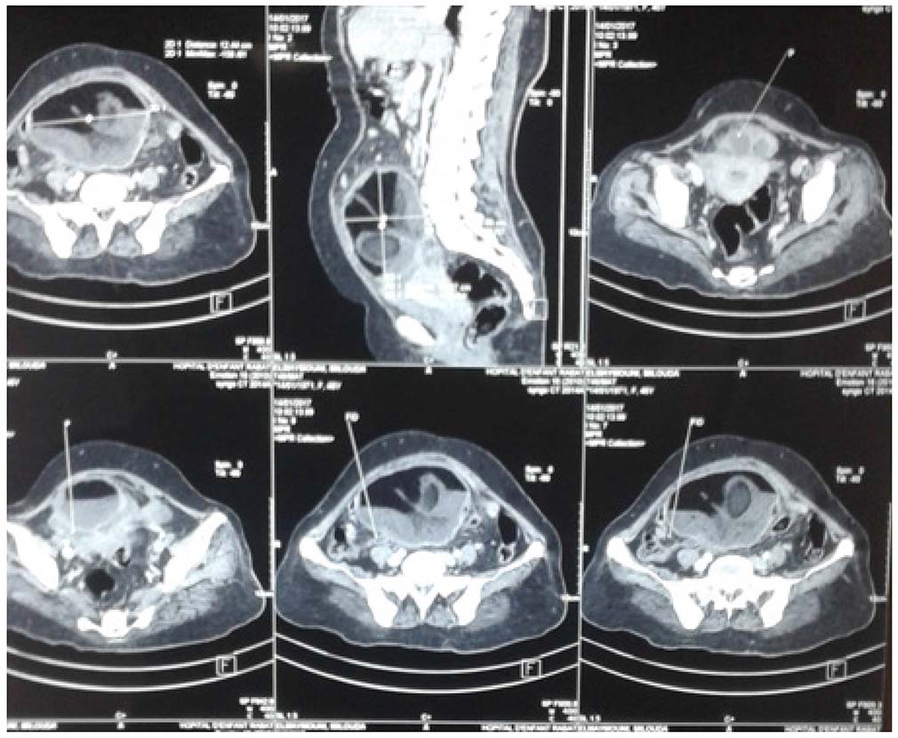
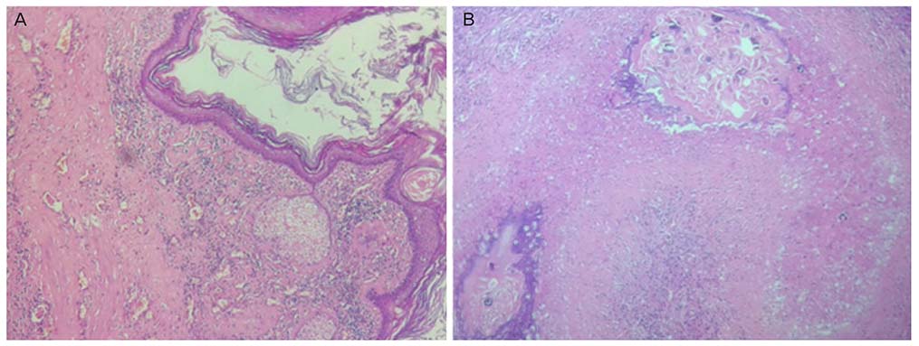
- Dermoid cysts or mature cystic teratoma are the most common type of ovarian germ cell tumor. It may be complicated by torsion, rupture, chemical peritonitis and malignant change but is rarely complicated by infection. We present a case of an ovarian dermoid cyst with super-infection caused by Schistosoma haematobium (S. haematobium). We present here a case of incidental finding of S. haematobium eggs in an infected cystic teratoma of the ovary because of the rare occurrence of this lesion. A 45-year-old Moroccan woman admitted to the gynecological department because of abdominal pain and fever. Gynecological examination, ultrasonography, and abdominopelvic computed tomography scan revealed an ovarian mass thought to be a dermoid cyst. The pathological evaluation suggested infected ovarian dermoid cyst with the presence of adult worm in the tumor, contains same eggs of S. haematobium. Super-infection of an ovarian dermoid cyst is a rare event, and the association with S. haematobium is extremely rare in the literature.
Keyword
MeSH Terms
Figure
Reference
-
1. Colley DG, Bustinduy AL, Secor WE, King CH. Human schistosomiasis. Lancet. 2014; 383:2253–2264.
Article2. Christinet V, Lazdins-Helds JK, Stothard JR, Reinhard-Rupp J. Female genital schistosomiasis (FGS): from case reports to a call for concerted action against this neglected gynecological disease. Int J Parasitol. 2016; 46:395–404.3. Abu Zikry AM, Fahmy K. Bilharziasis in a dermoid cyst of the ovary. J Obstet Gynaecol Br Commonw. 1963; 70:891–893.4. Paradinas FJ. Schistosomiasis in a cystic teratoma of the ovary. J Pathol. 1972; 106:123–126.
Article5. Sunder-Raj S. Cystic teratoma of ovary associated with schistosomiasis. East Afr Med J. 1976; 53:111–114.6. Melato M, Muuse MM, Hussein AM, Falconieri G. Schistosomiasis in a cystic teratoma of the ovary. Clin Exp Obstet Gynecol. 1987; 14:57–59.7. Sarma NH, Agnihotri S, Jeebun N. Incidental schistosomiasis in a dermoid cyst of the ovary: a case report. Internet J Parasit Dis. 2007; 3.
Article8. Hasanzadeh M, Tabare S, Mirzaean S. Ovarian dermoid cyst. Prof Med J. 2010; 17:512–515.9. Ayhan A, Bukulmez O, Genc C, Karamursel BS, Ayhan A. Mature cystic teratomas of the ovary: case series from one institution over 34 years. Eur J Obstet Gynecol Reprod Biol. 2000; 88:153–157.
Article10. Pradhan P, Thapa M. Dermoid cyst and its bizarre presentation. JNMA J Nepal Med Assoc. 2014; 52:837–844.
Article11. Chambô Filho A, Neves Ferreira R, Gusmão CB, Saade FT, Dalvi IR, Leo TC. Genital schistosomiasis: mucinous cystadenocarcinoma of the ovary containing schistosoma mansoni eggs. J Trop Med Parasitol. 2010; 33:36–40.12. Jourdan PM, Roald B, Poggensee G, Gundersen SG, Kjetland EF. Increased vascularity in cervicovaginal mucosa with Schistosoma haematobium infection. PLoS Negl Trop Dis. 2011; 5:e1170.13. Helling-Giese G, Sjaastad A, Poggensee G, Kjetland EF, Richter J, Chitsulo L, et al. Female genital schistosomiasis (FGS): relationship between gynecological and histopathological findings. Acta Trop. 1996; 62:257–267.
Article
- Full Text Links
- Actions
-
Cited
- CITED
-
- Close
- Share
- Similar articles
-
- A Case of Adenocarcinoma Arising from Dermoid Cyst of the Ovary
- A case of omental dermoid cyst due to an ovarian autoamputation
- A Case of Ovarian Dermoid Cyst Extending into the Urinary Bladder
- A Case of Congenital Limbal Dermoid Cyst
- Histopathologic Resemblance of Ovarian Dermoid Cyst to Various Skin Tumors


