Ann Hepatobiliary Pancreat Surg.
2019 Feb;23(1):84-86. 10.14701/ahbps.2019.23.1.84.
Intraoperatively malpositioned stent as a complication of common bile duct injury during laparoscopic cholecystectomy
- Affiliations
-
- 1Digestive Surgery Unit, Simone Veil General Hospital, Eaubonne, France. spleen2008@live.fr
- 2HPB Surgery Unit, Hammersmith Hospital, NHS/Imperial College, London, UK.
- 3HPB Surgery and Liver Transplantation Department, Beaujon Hospital, Clichy, France.
- 4Digestive Surgery and Surgical Oncology, Hôpital Avicenne, Assistance Publique - Hôpitaux de Paris, Paris, France.
- KMID: 2439053
- DOI: http://doi.org/10.14701/ahbps.2019.23.1.84
Abstract
- Injuries occurring during laparoscopic bile duct exploration in the course of laparoscopic cholecystectomy may represent threatening complications and lead to inappropriate management. We present a case of patient with biliary colic who underwent laparoscopic cholecystectomy. During the procedure, a common bile duct injury occurred, compelling conversion to open approach, and the patient was treated using a manually inserted biliary stent. She was referred with severe right upper quadrant pain six weeks after the surgery. Investigation with endoscopic retrograde cholangiopancreatography showed a malpositioned biliary stent with completely extra-biliary trajectory. This is thought to be the first description of a malpositioned common bile duct stent through the common biliary duct as a complication of the commonly performed surgical procedure of bile duct exploration.
Keyword
MeSH Terms
Figure
-
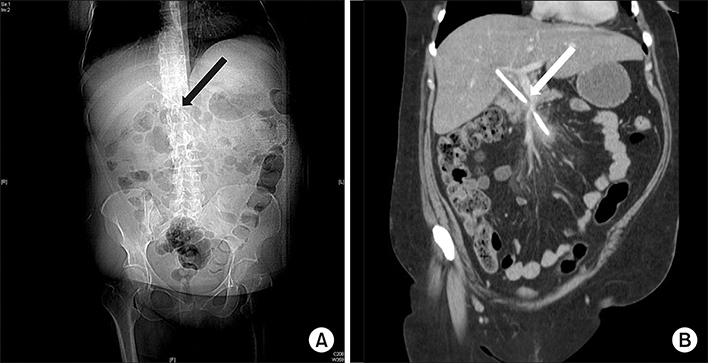
Fig. 1 X-ray and CT-scan. Imaging showing no abnormality regarding the abdominal X-ray (A) but a malpositioned biliary stent at abdominal CT-scan (B). The white arrow shows the biliary stent.
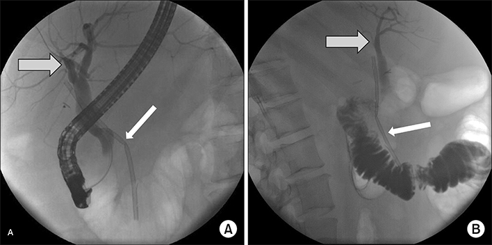
Fig. 2 Endoscopic Retrograde CholangioPancreatography. Endoscopic Retrograde cholangiopancreatography (A: front side; B: from the side) showing the biliary stent (white arrow) outside the dilated common bile duct (grey arrow).
Reference
-
1. Giger U, Ouaissi M, Schmitz SF, Krähenbühl S, Krähenbühl L. Bile duct injury and use of cholangiography during laparoscopic cholecystectomy. Br J Surg. 2011; 98:391–396.
Article2. Halbert C, Pagkratis S, Yang J, Meng Z, Altieri MS, Parikh P, et al. Beyond the learning curve: incidence of bile duct injuries following laparoscopic cholecystectomy normalize to open in the modern era. Surg Endosc. 2016; 30:2239–2243.
Article3. Way LW, Stewart L, Gantert W, Liu K, Lee CM, Whang K, et al. Causes and prevention of laparoscopic bile duct injuries: analysis of 252 cases from a human factors and cognitive psychology perspective. Ann Surg. 2003; 237:460–469.4. Sahajpal AK, Chow SC, Dixon E, Greig PD, Gallinger S, Wei AC. Bile duct injuries associated with laparoscopic cholecystectomy: timing of repair and long-term outcomes. Arch Surg. 2010; 145:757–763.5. Nicholson AA, Martin DF. Misplacement of endoscopic biliary endoprostheses. Endoscopy. 1997; 29:125–127.
Article6. Dumonceau JM, Tringali A, Blero D, Devière J, Laugiers R, Heresbach D, et al. Biliary stenting: indications, choice of stents and results: European Society of Gastrointestinal Endoscopy (ESGE) clinical guideline. Endoscopy. 2012; 44:277–298.
Article7. Strasberg SM. Error traps and vasculo-biliary injury in laparoscopic and open cholecystectomy. J Hepatobiliary Pancreat Surg. 2008; 15:284–292.
Article8. de Reuver PR, Grossmann I, Busch OR, Obertop H, van Gulik TM, Gouma DJ. Referral pattern and timing of repair are risk factors for complications after reconstructive surgery for bile duct injury. Ann Surg. 2007; 245:763–770.
Article
- Full Text Links
-
- Actions
-
Cited
- CITED
-
- Close
- Share
-
- Similar articles
-
- Recent classifications of the common bile duct injury
- Laparoscopic cholecystectomy and common bile duct exploration for gallstone and common bile duct stone in a patient with a left-sided gallbladder: a case report
- Surgical Clip Moved into the Extrahepatic Bile Duct after Laparoscopic Hepatectomy
- Clinical Experiences of Management of Bile Duct Injuries during Laparoscopic Cholecystectomy
- Relationship between the risk of bile duct injury during laparoscopic cholecystectomy and the types of preoperative magnetic resonance cholangiopancreatiocography (MRCP)
