Non-Operative Treatment for Symptomatic Osteochondral Lesion of the Talus
- Affiliations
-
- 1Department of Orthopedic Surgery, Asan Medical Center, University of Ulsan College of Medicine, Seoul, Korea. sgseo@amc.seoul.kr
- 2Department of Orthopedic Surgery, Gangneung Asan Hospital, University of Ulsan College of Medicine, Gangneung, Korea.
- KMID: 2438980
- DOI: http://doi.org/10.4055/jkoa.2017.52.2.153
Abstract
- PURPOSE
Although reports on operative treatment of osteochondral lesion of the talus (OLT) are increasing, to the best of our knowledge, there have been only a few reports on non-operative treatment of OLT. The purpose of this study is to report the prognosis of non-operative treatment for OLT patients.
MATERIALS AND METHODS
This retrospective study included 104 patients (57 male, 47 female) with OLTs having a follow-up period of more than two years, between 2003 and 2013. The location, size, and stage of the OLT were confirmed by magnetic resonance imaging or computed tomography. At the final follow-up, simple radiographs confirmed the progression of osteoarthritis. We surveyed the limitations of sports activity, visual analogue scale (VAS), American Orthopedic Foot and Ankle Society (AOFAS) scale, and SF-36.
RESULTS
There were no patients with progression of osteoarthritis at the final follow-up. Only two patients (2.4%) complained the limitation of desired sports activity. The mean VAS significantly decreased from 4.3 (range, 0-8) to 1.1 (range, 0-4) (p<0.001). The mean AOFAS scale significantly improved from 83.3 (range, 41-100) to 92.5 (range, 65-100). Moreover, the mean SF-36 also improved from 52.6 (range, 30.0-91.0) to 72.9 (range, 40.6-97.0) (p<0.001).
CONCLUSION
Sufficient non-operative treatment is initially recommended to OLT patients because pain, in general, improves in most cases despite the presence of symptoms. Moreover, it's worth noting that the progression to osteoarthritis is rare.
Keyword
MeSH Terms
Figure
-

Figure 1 Articular surface of talar dome is divided into nine areas.

Figure 2 Magnetic resonance imaging shows bone marrow edema and bone cyst of the talus.

Figure 3 An 18-year-old male. (A) Initial magnetic resonance imaging (MRI), (B) 10.4 years follow-up MRI. The size of the lesion decreased and American Orthopedic Foot and Ankle Society scale improved from 65 to 91.
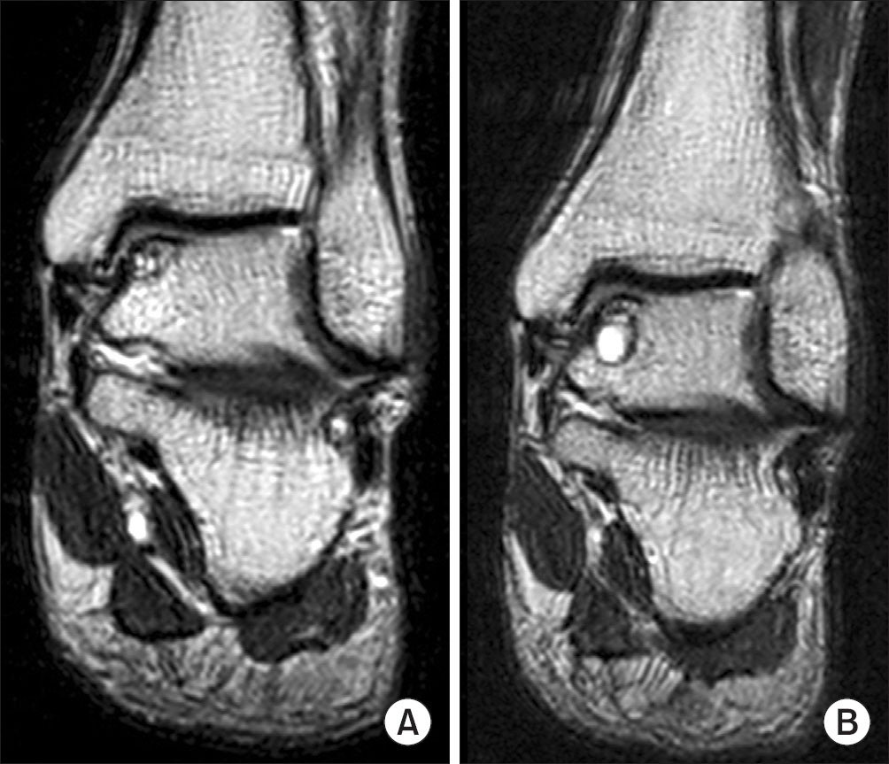
Figure 4 A 71-year-old male. (A) Initial magnetic resonance imaging (MRI), (B) 2.5 years follow-up MRI. The size of the lesion increased. However, American Orthopedic Foot and Ankle Society scale improved from 65 to 88.
Cited by 1 articles
-
Natural History of Osteochondral Lesion of the Talus
Min Gyu Kyung, Dong-Oh Lee, Dong Yeon Lee
J Korean Foot Ankle Soc. 2020;24(2):37-41. doi: 10.14193/jkfas.2020.24.2.37.
Reference
-
1. van Dijk CN, Reilingh ML, Zengerink M. van Bergen CJ. Osteochondral defects in the ankle: why painful? Knee Surg Sports Traumatol Arthrosc. 2010; 18:570–80.2. Bruns J. Osteochondrosis dissecans. Orthopade. 1997; 26:573–84.
Article3. Hannon CP, Smyth NA, Murawski CD. . Osteochondral lesions of the talus: aspects of current management. Bone Joint J. 2014; 96:164–71.4. Badekas T, Takvorian M, Souras N. Treatment principles for osteochondral lesions in foot and ankle. Int Orthop. 2013; 37:1697–706.
Article5. Murawski CD, Kennedy JG. Operative treatment of osteochondral lesions of the talus. J Bone Joint Surg Am. 2013; 95:1045–54.
Article6. Bauer M, Jonsson K, Lindén B. Osteochondritis dissecans of the ankle. A 20-year follow-up study. J Bone Joint Surg Br. 1987; 69:93–6.
Article7. Elias I, Jung JW, Raikin SM, Schweitzer MW, Carrino JA, Morrison WB. Osteochondral lesions of the talus: change in MRI findings over time in talar lesions without operative intervention and implications for staging systems. Foot Ankle Int. 2006; 27:157–66.
Article8. McCullough CJ, Venugopal V. Osteochondritis dissecans of the talus: the natural history. Clin Orthop Relat Res. 1979; 144:264–8.9. Pettine KA, Morrey BF. Osteochondral fractures of the talus. A long-term follow-up. J Bone Joint Surg Br. 1987; 69:89–92.
Article10. Shearer C, Loomer R, Clement D. Nonoperatively managed stage 5 osteochondral talar lesions. Foot Ankle Int. 2002; 23:651–4.
Article11. Takakura Y, Tanaka Y, Kumai T, Tamai S. Low tibialosteoto-my for osteoarthritis of the ankle. Results of a new operation in 18 patients. J Bone Joint Surg Br. 1995; 77:50–4.12. Davids JR, Gibson TW, Pugh LI. Quantitative segmental analysis of weight-bearing radiographs of the foot and ankle for children: normal alignment. J Pediatr Orthop. 2005; 25:769–76.13. Berndt AL, Harty M. Transchondral fractures (osteochondritis dissecans) of the talus. J Bone Joint Surg Am. 1959; 41:988–1020.
Article14. Ferkel RD. Scranton PE Jr. Arthroscopy of the ankle and foot. J Bone Joint Surg Am. 1993; 75:1233–42.15. Anderson IF, Crichton KJ, Grattan-Smith T, Cooper RA, Brazier D. Osteochondral fractures of the dome of the talus. J Bone Joint Surg Am. 1989; 71:1143–52.
Article16. Kitaoka HB, Alexander IJ, Adelaar RS, Nunley JA, Myer-son MS, Sanders M. Clinical rating systems for the ankle-hindfoot, midfoot, hallux, and lesser toes. Foot Ankle Int. 1994; 15:349–53.
Article17. Ware JE Jr, Sherbourne CD. The MOS 36-item short-form health survey (SF-36). I. Conceptual framework and item selection. Med Care. 1992; 30:473–83.18. Lee KM, Lee J, Chung CY. . Pitfalls and important issues in testing reliability using intraclass correlation coefficients in orthopaedic research. Clin Orthop Surg. 2012; 4:149–55.
Article19. Kumai T, Takakura Y, Higashiyama I, Tamai S. Arthroscopic drilling for the treatment of osteochondral lesions of the talus. J Bone Joint Surg Am. 1999; 81:1229–35.
Article20. Taranow WS, Bisignani GA, Towers JD, Conti SF. Retrograde drilling of osteochondral lesions of the medial talar dome. Foot Ankle Int. 1999; 20:474–80.
Article21. Klammer G, Maquieira GJ, Spahn S, Vigfusson V, Zanetti M, Espinosa N. Natural history of nonoperatively treated osteochondral lesions of the talus. Foot Ankle Int. 2015; 36:24–31.
Article
- Full Text Links
-
- Actions
-
Cited
- CITED
-
- Close
- Share
-
- Similar articles
-
- Autologous Osteochondral Transplantation as a Secondary Procedure after Failed Microfracture for Osteochondral Lesion of Talus
- Simultaneously Occurred Medial and Lateral Osteochondral Lesions of the Talus
- Redomicrofracture as a Treatment for Osteochondral Lesion of Talus after the Failure of Arthroscopic Microfracture
- Osteochondral Lesion of the Talus in Children and Adolescents
- Autologous Chondrocyte Implantation as a Secondary Procedure after Failed Microfracture for Osteochondral Lesion of Talus
