Papillary Cannulation Facilitated by Submucosal Saline Injection into an Intradiverticular Papilla
- Affiliations
-
- 1Department of Internal Medicine, Chungbuk National University Hospital, Cheongju, Korea. smpark@chungbuk.ac.kr
- 2Department of Internal Medicine, Chungbuk National University College of Medicine, Cheongju, Korea.
- KMID: 2438151
- DOI: http://doi.org/10.5946/ce.2018.060
Abstract
- Endoscopic retrograde cholangiopancreatography (ERCP) of the intradiverticular papilla with its invisible orifice remains challenging. Several techniques have been introduced to evert the papillary opening to facilitate cannulation. A 79-year-old woman with bile duct stones underwent ERCP, which revealed that the papilla was located inside a large diverticulum and tended to rotate inward with a trial of papillary cannulation. Submucosal papillary injection of 3 cc of normal saline was performed at 3 and 9 o'clock. Eversion and fixation of a papilla in the diverticulum with this technique allowed selective cannulation of the biliary tree. Stones were retrieved after endoscopic papillary balloon dilation without complications. She had an uneventful post-procedural course. Our findings suggest that submucosal saline injection technique is safe and effective for selective cannulation and can be recommended when cannulation is very difficult because of an intradiverticular papilla.
Keyword
MeSH Terms
Figure
-
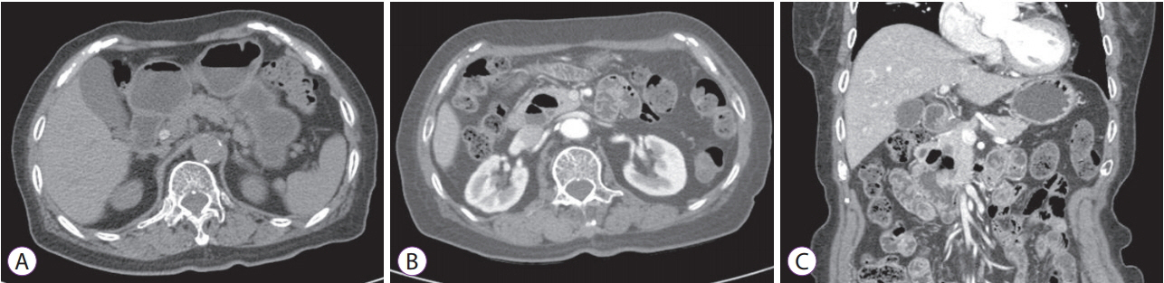
Fig. 1. Abdominal computed tomography. (A) Pre-contrast axial scan showed a hyperdense lesion inside the dilated common bile duct. (B, C) Post-contrast axial scan and coronal view revealed the presence of a periampullary diverticulum (arrowheads).
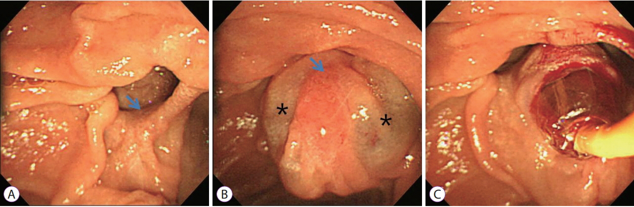
Fig. 2. Duodenoscopic findings during endoscopic retrograde cholangiopancreatography. (A) The major duodenal papilla (arrow) is positioned inside the diverticulum. (B) Saline injection was performed at 3 and 9 o’clock (stars) for eversion and fixation of the papilla (arrow). (C) Endoscopic papillary balloon dilation preceded stone extraction.
Cited by 1 articles
-
A “One Accessory and One Guidewire-in-One Channel” Technique in a Patient with Billroth II Anastomosis
Kook Hyun Kim, Sung Bum Kim, Tae Nyeun Kim
Clin Endosc. 2021;54(1):139-140. doi: 10.5946/ce.2020.087.
Reference
-
1. Fogel EL, Sherman S, Lehman GA. Increased selective biliary cannulation rates in the setting of periampullary diverticula: main pancreatic duct stent placement followed by pre-cut biliary sphincterotomy. Gastrointest Endosc. 1998; 47:396–400.
Article2. Parlak E, Suna N, Kuzu UB, et al. Diverticulum with papillae: does position of papilla affect technical success? Surg Laparosc Endosc Percutan Tech. 2015; 25:395–398.3. Boix J, Lorenzo-Zúñiga V, Añaños F, Domènech E, Morillas RM, Gassull MA. Impact of periampullary duodenal diverticula at endoscopic retrograde cholangiopancreatography: a proposed classification of periampullary duodenal diverticula. Surg Laparosc Endosc Percutan Tech. 2006; 16:208–211.
Article4. Chen L, Xia L, Lu Y, Bie L, Gong B. Influence of periampullary diverticulum on the occurrence of pancreaticobiliary diseases and outcomes of endoscopic retrograde cholangiopancreatography. Eur J Gastroenterol Hepatol. 2017; 29:105–111.
Article5. Güitrón-Cantú A, Adalid-Martínez R, Gutiérrez-Bermudez JA, Segura-López FK, García-Vázquez A. [Difficulty to cannulate papilla of Vater in the presence of periampullary duodenal diverticulum]. Rev Gastroenterol Mex. 2010; 75:273–280.6. Harada H, Suehiro S, Shimizu T, Katsuyama Y, Hayasaka K. Submucosal injection can facilitate biliary access in patients with periampullary diverticula. Gastrointest Endosc. 2016; 84:185–186.
Article7. Altonbary AY, Bahgat MH. Endoscopic retrograde cholangiopancreatography in periampullary diverticulum: the challenge of cannulation. World J Gastrointest Endosc. 2016; 8:282–287.
Article8. Parlak E, Köksal AS, Dişibeyaz S, et al. Additional biliary cannulation methods in patients with juxtapapillary duodenal diverticula. Dig Dis Sci. 2012; 57:2982–2987.
Article9. Cappell MS, Mogrovejo E, Manickam P, Batke M. Endoclips to facilitate cannulation and sphincterotomy during ERCP in a patient with an ampulla within a large duodenal diverticulum: case report and literature review. Dig Dis Sci. 2015; 60:168–173.
Article10. Fujita N, Noda Y, Kobayashi G, Kimura K, Yago A. ERCP for intradiverticular papilla: two-devices-in-one-channel method. Endoscopic retrograde cholangiopancreatography. Gastrointest Endosc. 1998; 48:517–520.11. Elmunzer BJ, Boetticher NC. Reverse guidewire anchoring of the papilla for difficult cannulation due to a periampullary diverticulum. Gastrointest Endosc. 2015; 82:957.
Article12. Külling D, Haskell E. Double endoscope method to access intradiverticular papilla. Gastrointest Endosc. 2005; 62:811–812.
Article13. Myung DS, Park CH, Koh HR, et al. Cap-assisted ERCP in patients with difficult cannulation due to periampullary diverticulum. Endoscopy. 2014; 46:352–355.
Article14. Wang BC, Shi WB, Zhang WJ, et al. Entering the duodenal diverticulum: a method for cannulation of the intradiverticular papilla. World J Gastroenterol. 2012; 18:7394–7396.
Article15. Kim HJ, Kim YS, Myung SJ, et al. A novel approach for cannulation to the ampulla within the diverticulum: double-catheter method. Endoscopy. 1998; 30:S103–S104.
Article16. Tóth E, Lindström E, Fork FT. An alternative approach to the inaccessible intradiverticular papilla. Endoscopy. 1999; 31:554–556.
Article17. Calvo MM, Bujanda L, Heras I, et al. The rendezvous technique for the treatment of choledocholithiasis. Gastrointest Endosc. 2001; 54:511–513.
Article18. Tarantino I, Barresi L, Fabbri C, Traina M. Endoscopic ultrasound guided biliary drainage. World J Gastrointest Endosc. 2012; 4:306–311.
Article19. Testoni PA, Mariani A, Aabakken L, et al. Papillary cannulation and sphincterotomy techniques at ERCP: European Society of Gastrointestinal Endoscopy (ESGE) clinical guideline. Endoscopy. 2016; 48:657–683.
Article20. García-Cano J. Use of an ultrathin gastroscope to locate a papilla hidden within a duodenal diverticulum. Endoscopy. 2010; 42 Suppl 2:E96–E97.
Article
- Full Text Links
-
- Actions
-
Cited
- CITED
-
- Close
- Share
-
- Similar articles
-
- Endoclip-Assisted Cannulation for a Hidden Duodenal Papilla: Three Cases
- Factors Predicting Difficult Biliary Cannulation during Endoscopic Retrograde Cholangiopancreatography for Common Bile Duct Stones
- Clinical Characteristics and Endoscopic Findings of Impacted Papillary Stone
- Application of Grasp Forceps on Selective Cannulation of Pancreatobiliary Duct in a Patient with Peripapillary Diverticulum
- The Application of Clips during ERCP: A New Anchoring Method for Redundant Kerckrings Folds Covering the Duodenal Papilla
