J Korean Ophthalmol Soc.
2019 Feb;60(2):181-186. 10.3341/jkos.2019.60.2.181.
Factors Affecting Persistent Diplopia after Surgical Repair of Isolated Inferior Orbital Wall Fracture
- Affiliations
-
- 1Department of Ophthalmology, Inha University School of Medicine, Incheon, Korea. ksm0724@inha.ac.kr
- KMID: 2438078
- DOI: http://doi.org/10.3341/jkos.2019.60.2.181
Abstract
- PURPOSE
To investigate factors affecting persistent diplopia after surgical repair of isolated inferior orbital wall fractures.
METHODS
Thirty-three patients who underwent surgical repair of isolated inferior orbital wall fractures in Inha University Hospital Ophthalmology Department from 2014 to 2017 were enrolled in this study. The authors examined facial computed tomography, diplopia, extraocular muscle movement, and Hertel's exophthalmometer before and 6 months after surgery. The diplopia which was not recovered even at 6 months postoperatively was defined as persistent diplopia. Multivariable logistic regression analyses were performed on parameters that were found to be related to persistent diplopia using univariable logistic regression analyses.
RESULTS
Univariable regression analysis showed that preoperative ocular motility limitation, preoperative diplopia, the type of fracture, the number of contacts with the fracture site and extraocular muscle (EOM), and EOM tenting were associated with persistent postoperative diplopia. Multivariable regression analysis using the previously mentioned five parameters showed 28.3-fold and 17.4-fold greater probabilities of diplopia after surgery in preoperative diplopia and EOM tenting, respectively (p = 0.023).
CONCLUSIONS
Preoperative diplopia and EOM tenting were associated with persistent postoperative diplopia. These parameters were predictors of persistent diplopia in eyes with isolated inferior orbital wall fractures.
Keyword
Figure
-
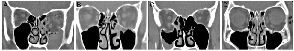
Figure 1 Fracture type. (A) Open type, extraocular muscle (EOM) displacement, EOM deformity. (B) Close type, EOM tenting (focal angulation). (C) Open type, EOM tenting (focal angulation), contact between EOM and bony edge. (D) Close type, EOM swelling.
Reference
-
1. Dodick JM, Galin MA, Littleton JT, Sod LM. Concomitant medial wall fracture and blowout fracture of the orbit. Arch Ophthalmol. 1971; 85:273–276.2. Dulley B, Fells P. Long-term follow-up of orbital blow-out fractures with and without surgery. Mod Probl Ophthalmol. 1975; 14:467–470.3. Davidson TM, Olesen RM, Nahum AM. Medial orbital wall fracture with rectus entrapment. Arch Otolaryngol. 1975; 101:33–35.
Article4. Cruz AA, Eichenberger GC. Epidemiology and management of orbital fractures. Curr Opin Ophthalmol. 2004; 15:416–421.
Article5. Harris GJ. Orbital blow-out fractures: surgical timing and technique. Eye (Lond). 2006; 20:1207–1212.
Article6. Hoşal BM, Beatty RL. Diplopia and enopthalmos after surgical repair of blowout fracture. Orbit. 2002; 21:27–33.7. Putterman AM, Stevens T, Urist MJ. Nonsurgical management of blow-out fractures of the orbital floor. Am J Ophthalmol. 1974; 77:232–239.
Article8. Gilbard SM, Mafee MF, Lagouros PA, Langer BG. Orbital blowout fractures. The prognostic significance of computed tomography. Ophthalmology. 1985; 92:1523–1528.
Article9. Pearl RM. Treatment of enopthalmos. Clin Plast Surg. 1992; 19:99–111.10. Iliff NT. The ophthalmic implications of the correction of late enophthalmos following severe midfacial trauma. Trans Am Ophthalmol Soc. 1991; 89:477–548.11. Hwang JH, Kwak MS. Residual diplopia and enophthalmos after reconstruction of orbital wall fractures. J Korean Ophthalmol Soc. 2003; 44:1959–1965.12. Jung H, Byun JY, Kim HJ, et al. Prognostic CT findings of diplopia after surgical repair of pure orbital blowout fracture. J Craniomaxillofac Surg. 2016; 44:1479–1484.
Article13. Harris GJ, Garcia GH, Logani SC, Murphy ML. Correlation of preoperative computed tomography and postoperative ocular motility in orbital blowout fractures. Ophthalmic Plast Reconstr Surg. 2000; 16:179–187.
Article14. Jin HR, Lee HS, Yeon JY, Suh MW. Residual diplopia after repair of pure orbital blowout fracture: the importance of extraocular muscle injury. Am J Rhinol. 2007; 21:276–280.
Article15. Yano H, Suzuki Y, Yoshimoto H, et al. Linear-type orbital floor fracture with or without muscle involvement. J Craniofac Surg. 2010; 21:1072–1078.
Article16. Lee SY, Kim SY, Kim HB. Orbital fractures evaluated by computed tomography. J Korean Ophthalmol Soc. 1990; 31:249–253.17. Ahn SK, Jung SW. The clinical aspects of orbital fractures proven by computed tomography. J Korean Ophthalmol Soc. 1997; 38:2077–2083.18. Tahiri Y, Lee J, Tahiri M, et al. Preoperative diplopia: the most important prognostic factor for diplopia after surgical repair of pure orbital blowout fracture. J Craniofac Surg. 2010; 21:1038–1041.19. Matsunaga K, Asamura S, Morotomi T, et al. Association between preoperative inferior rectus muscle swelling and outcomes in orbital blowout fracture. J Craniomaxillofac Surg. 2011; 39:509–514.
Article20. Furuta M, Yago K, Iida T. Correlation between ocular motility and evaluation of computed tomography in orbital blowout fracture. Am J Ophthalmol. 2006; 142:1019–1025.
Article21. Kang HJ, Ha MS. A clinical feature of the patients of orbital wall fracture with diplopia. J Korean Ophthalmol Soc. 2009; 50:969–975.
Article22. Hawes MJ, Dortzbach RK. Surgery on orbital floor fractures. Influence of time of repair and fracture size. Ophthalmology. 1983; 90:1066–1070.23. Jang KH, Kim NJ, Choung HK, Khwarg SI. Orbital wall fracture repair: the results of early and delayed surgery. J Korean Ophthalmol Soc. 2016; 57:181–187.
Article24. Koornneef L. Orbital septa: anatomy and function. Ophthalmology. 1979; 86:876–878.
Article25. Koornneef L. Current concepts on the management of orbital blow-out fractures. Ann Plast Surg. 1982; 9:185–200.
Article26. Koornneef L, Zonneveld FW. The role of direct multiplanar high resolution CT in the assessment and management of orbital trauma. Radiol Clin North Am. 1987; 25:753–766.27. Hopper RA, Salemy S, Sze RW. Diagnosis of midface fractures with CT: what the surgeon needs to know. Radiographics. 2006; 26:783–793.
Article
- Full Text Links
-
- Actions
-
Cited
- CITED
-
- Close
- Share
-
- Similar articles
-
- Diplopia after Isolated Inferior Orbital Wall Fracture According to the Computed Tomography Findings
- The Clinical Aspects of Orbital Fractures Proven by Computed Tomography
- Residual Diplopia and Enophthalmos after Reconstruction of Orbital Wall Fractures
- Repair of Medial Orbital Wall Fracture
- The Inferior Orbital Wall Reconstruction by Titanium Micro-mesh Remodeling
