RASA1-Related Parkes Weber Syndrome in a Neonate
- Affiliations
-
- 1Department of Pediatrics, Cheil General Hospital & Women's Healthcare Center, Dankook University College of Medicine, Seoul, Korea. ykleeped@hanmail.net
- 2Department of Radiology, Cheil General Hospital & Women's Healthcare Center, Dankook University College of Medicine, Seoul, Korea.
- KMID: 2436129
- DOI: http://doi.org/10.5385/nm.2018.25.3.126
Abstract
- Parkes Weber syndrome is a rare congenital vascular anomaly, related to the RAS p21 protein activator 1 (RASA1) gene. It is characterized by capillary cutaneous malformations, bony and soft tissue hyperplasia, and multiple arteriovenous fistulas throughout the affected upper or lower extremity. These arteriovenous fistulas can be associated with life-threatening complications such as bleeding, thrombosis, and high output heart failure. In this report, we present a neonate who had a disproportionately hypertrophied left upper limb with port-wine stain, dystrophy of the left humerus, and hypertrophy of the left clavicle on X-ray, and arteriovenous malformation and massive dilatation of the left subclavian artery on magnetic resonance angiography. Exome sequencing analysis revealed a novel heterozygous splicing mutation (c.1776+2T>A) in the RASA1 gene. To the best of our knowledge, this report is the first case of RASA1-related Parkes Weber syndrome in Korea.
Keyword
MeSH Terms
Figure
-
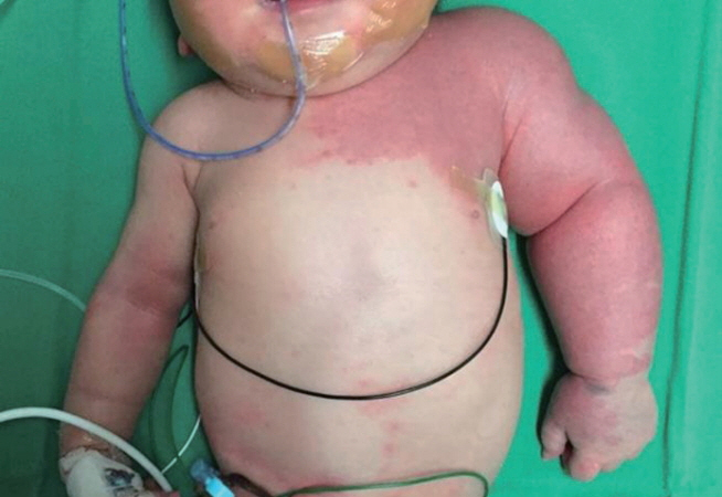
Figure 1. Photograph showing an asymmetric hypertrophy of the left upper extremity with an extensive geographic port-wine stain.
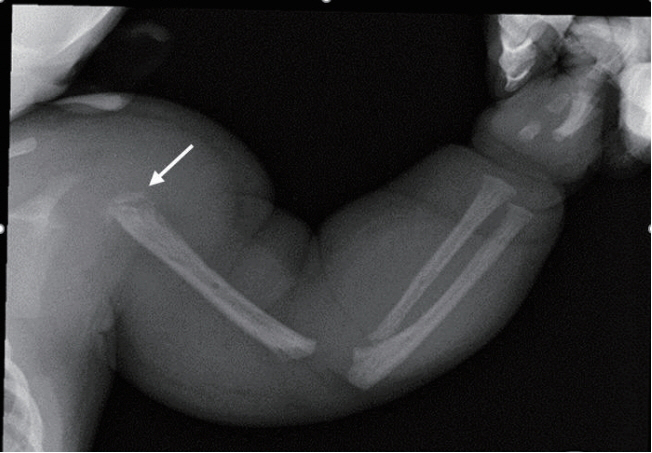
Figure 2. Radiograph of the left arm shows dystrophy of the left humerus (arrow) and diffuse soft tissue swelling of the left arm and shoulder.
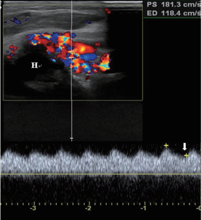
Figure 3. Doppler ultrasound of the left arm demonstrates a high velocity waveform (181.3 cm/sec) with increased diastolic flow (arrow) due to an arteriovenous shunt in the upper arm around the humeral head (H).

Figure 4. Magnetic resonance angiogram of the left arm shows engorged left subclavian, and axillary arteries (arrow) with their branches including left circumflex humeral arteries, arteries of forearm, and muscular branches (arrowhead).

Figure 5. Chromatogram of the patient (proband) and family members for the RAS p21 protein activator 1 (RASA1) gene. Genetic analysis showing the sequence of exon 13 and intron 13 of RASA1, where the mutation was detected in the patient. The mutation is c.1776+2T>A. His parents and brother had no mutation of the gene.
Reference
-
1. Parkes Weber F, Liang MG. Angioma: formation in connection with hypertrophy of limbs and hemi-hypertrophy. Br J Dermatol. 1907; 19:231–5.2. Maguiness SM, Liang MG. Management of capillary malformations. Clin Plast Surg. 2011; 38:65–73.3. Nozaki T, Nosaka S, Miyazaki O, Makidono A, Yamamoto A, Niwa T, et al. Syndromes associated with vascular tumors and malformations: a pictorial review. Radiographics. 2013; 33:175–95.4. Enjolras O, Chapot R, Merland JJ. Vascular anomalies and the growth of limbs: a review. J Pediatr Orthop B. 2004; 13:349–57.5. Eerola I, Boon LM, Mulliken JB, Burrows PE, Dompmartin A, Watanabe S, et al. Capillary malformation-arteriovenous malformation, a new clinical and genetic disorder caused by RASA1 mutations. Am J Hum Genet. 2003; 73:1240–9.6. Gloviczki P, Duncan A, Kalra M, Oderich G, Ricotta J, Bower T, et al. Vascular malformations: an update. Perspect Vasc Surg Endovasc Ther. 2009; 21:133–48.7. Adekanmi AJ, Schernthaner RE, Lammer J. Parkes Weber syndrome: a rare vascular malformation-imaging and the usefulness of intravascular intervention. Internet J Radiol. 2015; 18:1–7. https://doi.org/10.5580/IJRA.24813.8. Hershkovitz D, Bergman R, Sprecher E. A novel mutation in RASA1 causes capillary malformation and limb enlargement. Arch Dermatol Res. 2008; 300:385–8.9. Revencu N, Boon LM, Mulliken JB, Enjolras O, Cordisco MR, Burrows PE, et al. Parkes Weber syndrome, vein of Galen aneurysmal malformation, and other fast-flow vascular anomalies are caused by RASA1 mutations. Hum Mutat. 2008; 29:959–65.10. Behr GG, Liberman L, Compton J, Garzon MC, Morel KD, Lauren CT, et al. CM-AVM syndrome in a neonate: case report and treatment with a novel flow reduction strategy. Vasc Cell. 2012; 4:19.11. Banzic I, Brankovic M, Maksimovic Z, Davidovic L, Markovic M, Rancic Z. Parkes Weber syndrome-diagnostic and management paradigms: a systematic review. Phlebology. 2017; 32:371–83.12. Ziyeh S, Spreer J, Rossler J, Strecker R, Hochmuth A, Schumacher M, et al. Parkes Weber or Klippel-Trenaunay syndrome? Non-invasive diagnosis with MR projection angiography. Eur Radiol. 2004; 14:2025–9.13. Jamis-Dow CA, Turner J, Biesecker LG, Choyke PL. Radiologic manifestations of Proteus syndrome. Radiographics. 2004; 24:1051–68.14. Paltiel HJ, Burrows PE, Kozakewich HP, Zurakowski D, Mulliken JB. Soft-tissue vascular anomalies: utility of US for diagnosis. Radiology. 2000; 214:747–54.15. Lobo-Mueller E, Amaral JG, Babyn PS, Wang Q, John P. Complex combined vascular malformations and vascular malformation syndromes affecting the extremities in children. Semin Musculoskelet Radiol. 2009; 13:255–76.16. Kulkarni SV, Gish G, van der Geer P, Henkemeyer M, Pawson T. Role of p120 Ras-GAP in directed cell movement. J Cell Biol. 2000; 149:457–70.17. Triana P, Dore M, Cerezo VN, Cervantes M, Sanchez AV, Ferrero MM, et al. Sirolimus in the treatment of vascular anomalies. Eur J Pediatr Surg. 2017; 27:86–90.
