Ann Dermatol.
2012 May;24(2):240-241.
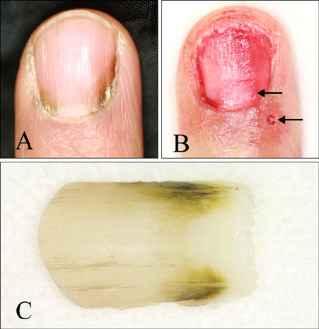
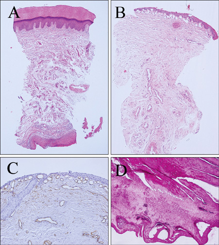
Proximal and Lateral Chromonychia with Capillary Proliferation on the Distal Nail Matrix
- Affiliations
-
- 1Department of Dermatology, Yeouido St. Mary's Hospital, College of Medicine, The Catholic University of Korea, Seoul, Korea. hjpark@catholic.ac.kr
Abstract
- No abstract available.
MeSH Terms
Figure
Reference
-
1. Baran R, Dawber RPR. Baran and Dawber's diseases of the nails and their management. Malden: Blackwell Science;85–86.2. Tosti A, Daniel CR, Piraccini BM, Lorizzo M. Color atlas of nails. 2010. New York: Springer;45–60.3. Olsen TG, Jatlow P. Contact exposure to elemental iron causing chromonychia. Arch Dermatol. 1984. 120:102–103.
Article4. Roh M, Lee J, Lee K. A case of chromonychia with hyperbilirubinemia. J Eur Acad Dermatol Venereol. 2007. 21:127–128.
Article
- Full Text Links
- Actions
-
Cited
- CITED
-
- Close
- Share
- Similar articles
-
- A Case of Chronic Longitudinal Hemosiderin Chromonychia with Nail Plate Deformities Mimicking Nail Malignancy
- A Case of Multiple Periungual Fibrokeratoma with Matrix Differentiation
- Problems in Humeral Interlocking with Seidel Nail
- 1,064-nm and 532-nm picosecond neodymium-doped:yttrium-aluminum-garnet laser treatment for longitudinal melanonychia: a case report
- Immediate Nail Bed Graft on Exposed Distal Phalanx in Fingertip Injury


