Ann Dermatol.
2012 May;24(2):220-222.
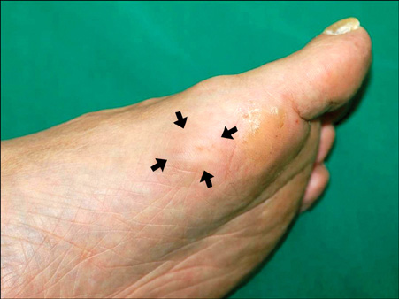
Solitary Cutaneous Myofibroma on the Sole: An Unusual Localization
- Affiliations
-
- 1Department of Dermatology, Hanyang University Guri Hospital, Hanyang University College of Medicine, Guri, Korea. tuentuen@hanyang.ac.kr
Abstract
- No abstract available.
MeSH Terms
Figure
Reference
-
1. Holst VA, Junkins-Hopkins JM, Elenitsas R. Cutaneous smooth muscle neoplasms: clinical features, histologic findings, and treatment options. J Am Acad Dermatol. 2002. 46:477–490.
Article2. Guitart J, Ritter JH, Wick MR. Solitary cutaneous myofibromas in adults: report of six cases and discussion of differential diagnosis. J Cutan Pathol. 1996. 23:437–444.
Article3. Beham A, Badve S, Suster S, Fletcher CD. Solitary myofibroma in adults: clinicopathological analysis of a series. Histopathology. 1993. 22:335–341.
Article4. Requena L, Kutzner H, Hügel H, Rütten A, Furio V. Cutaneous adult myofibroma: a vascular neoplasm. J Cutan Pathol. 1996. 23:445–457.
Article5. Chang JY, Kessler HP. Masson trichrome stain helps differentiate myofibroma from smooth muscle lesions in the head and neck region. J Formos Med Assoc. 2008. 107:767–773.
Article


