J Korean Fract Soc.
2019 Jan;32(1):56-60. 10.12671/jkfs.2019.32.1.56.
Bilateral Gluteal Necrosis and Deep Infection after Transarterial Embolization for Pelvic Ring Injury in Patient with Hemodynamic Instability: A Case Report
- Affiliations
-
- 1Department of Trauma Surgery, Pusan National University Hospital, Busan, Korea.
- 2Department of Intervention Radiology, Pusan National University Hospital, Busan, Korea.
- 3Department of Orthopaedic Surgery, Pusan National University Hospital, Busan, Korea. realjang1979@gmail.com
- KMID: 2432500
- DOI: http://doi.org/10.12671/jkfs.2019.32.1.56
Abstract
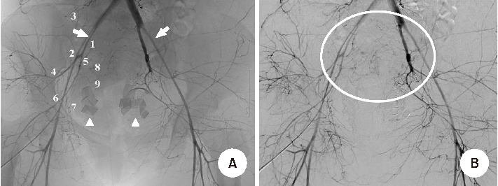
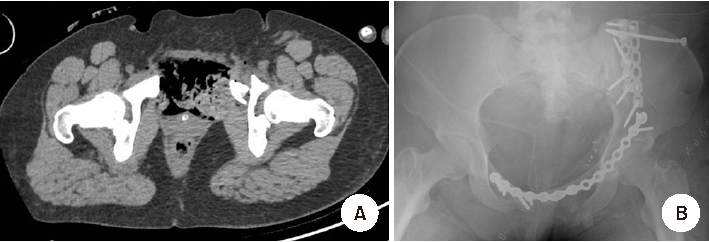
- Transarterial embolization is accepted as effective and safe for the acute management in hemodynamically unstable patients with pelvic ring injury. However, transarterial embolization has potential complications, such as gluteal muscle/skin necrosis, deep infection, surgical wound breakdown, and internal organ infarction, which are caused by blocked blood flow to surrounding tissues and organs, and many studies on the complications have been reported. Here, we report an experience of the management of gluteal necrosis and infection that occurred after transarterial embolization, with a review of the relevant literature.
Figure
Reference
-
1. El-Haj M, Bloom A, Mosheiff R, Liebergall M, Weil YA. Outcome of angiographic embolisation for unstable pelvic ring injuries: factors predicting success. Injury. 2013; 44:1750–1755.
Article2. Lustenberger T, Wutzler S, Störmann P, Laurer H, Marzi I. The role of angio-embolization in the acute treatment concept of severe pelvic ring injuries. Injury. 2015; 46:Suppl 4. S33–S38.
Article3. Perez JV, Hughes TM, Bowers K. Angiographic embolisation in pelvic fracture. Injury. 1998; 29:187–191.
Article4. Takahira N, Shindo M, Tanaka K, Nishimaki H, Ohwada T, Itoman M. Gluteal muscle necrosis following transcatheter angiographic embolisation for retroperitoneal haemorrhage associated with pelvic fracture. Injury. 2001; 32:27–32.
Article5. Matityahu A, Marmor M, Elson JK, et al. Acute complications of patients with pelvic fractures after pelvic angiographic embolization. Clin Orthop Relat Res. 2013; 471:2906–2911.
Article6. Tanizaki S, Maeda S, Matano H, Sera M, Nagai H, Ishida H. Time to pelvic embolization for hemodynamically unstable pelvic fractures may affect the survival for delays up to 60 min. Injury. 2014; 45:738–741.
Article7. Velmahos GC, Chahwan S, Falabella A, Hanks SE, Demetriades D. Angiographic embolization for intraperitoneal and retroperitoneal injuries. World J Surg. 2000; 24:539–545.
Article8. Li Q, Dong J, Yang Y, et al. Retroperitoneal packing or angioembolization for haemorrhage control of pelvic fractures--Quasi-randomized clinical trial of 56 haemodynamically unstable patients with Injury Severity Score ≥33. Injury. 2016; 47:395–401.
Article9. Yoon HK, Kim MD, Han SH, Kim BK, Ahn TK. Effectiveness of arterial embolization in hemodynamically unstable pelvic fracture. J Korean Hip Soc. 2008; 20:117–123.
Article10. Hak DJ, Smith WR, Suzuki T. Management of hemorrhage in life-threatening pelvic fracture. J Am Acad Orthop Surg. 2009; 17:447–457.
Article
- Full Text Links
- Actions
-
Cited
- CITED
-
- Close
- Share
- Similar articles
-
- Superior Gluteal Artery Injury during Percutaneous Iliosacral Screw Fixation: A Case Report
- Early Results of Percutaneous Ilioscral Screw Fixation in Unstable Posterior Pelvic Ring Injury
- Latent Superior Gluteal Artery Injury by Entrapment between the Fragments in Transverse Acetabular Fracture - A Case Report -
- Testicular Dislocation Associated with Pelvic Ring Injury
- A Case of Uterine Fibroids Necrosis after Transarterial Embolization for Treatment of Uterine Fibroids


