Korean Circ J.
2019 Feb;49(2):194-196. 10.4070/kcj.2018.0309.
Sinus of Valsalva Aneurysm and Multiple Aortic Aneurysms Provoked by Viral Myocarditis
- Affiliations
-
- 1Department of Cardiology, Ajou University School of Medicine, Suwon, Korea. lavioli@hanmail.net
- KMID: 2432139
- DOI: http://doi.org/10.4070/kcj.2018.0309
Abstract
- No abstract available.
Figure
-
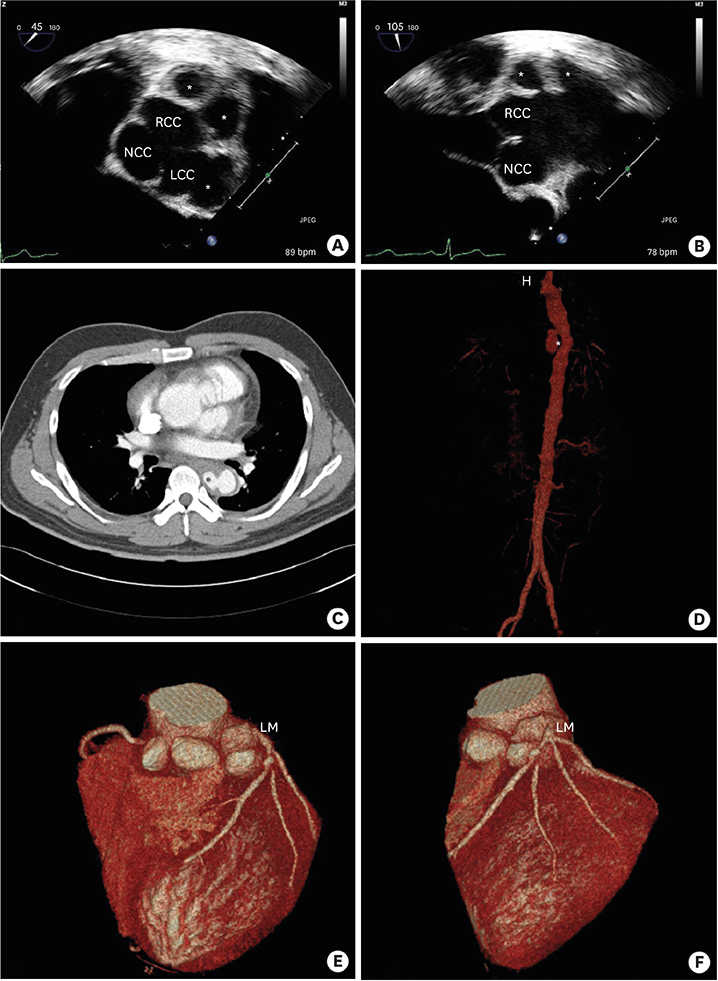
Figure 1 (A) Transesophageal echocardiography revealed that the outpouching structure with mural thrombus arose from the left coronary sinus of Valsalva. (A and B) Distal to the right coronary ostium, the aorta formed another 2 outpouching structures. (C and D) CT revealed another outpouching structure at descending aorta. (E and F) Cardiac multi-detector CT revealed that the left coronary ostium was originated from the aneurismal wall. CT = computed tomography; LCC = left coronary cusp; LM = left main artery; NCC = non-coronary cusp; RCC = right coronary cusp. *Aneurysm.
Reference
-
1. Batiste C, Bansal RC, Razzouk AJ. Echocardiographic features of an unruptured mycotic aneurysm of the right aortic sinus of Valsalva. J Am Soc Echocardiogr. 2004; 17:474–477.
Article2. Medeiros Sobrinho JH, Silva MA, Fontes WF, et al. Syphilitic aneurysm communicating with an aortic sinus of Valsalva. A case report. Arq Bras Cardiol. 1989; 52:341–344.
- Full Text Links
-
- Actions
-
Cited
- CITED
-
- Close
- Share
-
- Similar articles
-
- Surgical Correction of Ruptured Aneurysm of Aortic Sinus of Valsalva
- Two cases of ruptured congenital sinus of Valsalva aneurysms dissecting into the interventricular septum in patients with cerebral infarction
- A Case of Perimembranous Ventricular Septal Defect Associated with Sinus of Valsalva Aneurysm Mimicking Membranous Septal Aneurysm
- Simultaneous Aortic and Tricuspid Valve Endocarditis due to Complication of Sinus of Valsalva Rupture
- Extracardiac Aneurysm of the Sinus of Valsalva: A Case Report
