Korean Circ J.
2019 Feb;49(2):173-180. 10.4070/kcj.2018.0152.
Benefit of Four-Dimensional Computed Tomography Derived Ejection Fraction of the Left Atrial Appendage to Predict Thromboembolic Risk in the Patients with Valvular Heart Disease
- Affiliations
-
- 1Division of Cardiology, Severance Cardiovascular Hospital, Yonsei University College of Medicine, Seoul, Korea. HJCHANG@yuhs.ac
- 2Division of Cardiology, Department of Internal Medicine, Dongsan Medical Center, Keimyung University College of Medicine, Daegu, Korea.
- 3Department of Neurology, Yonsei University College of Medicine, Seoul, Korea.
- 4Department of Radiology, Severance Hospital, Yonsei University College of Medicine, Seoul, Korea.
- KMID: 2432135
- DOI: http://doi.org/10.4070/kcj.2018.0152
Abstract
- BACKGROUND AND OBJECTIVES
Decreased left atrial appendage (LAA) emptying velocity in transesophageal echocardiography (TEE) is related with higher incidence of thrombus and increased risk of stroke. Patients with valve disease are at higher risk of thrombus formation before and after surgery. The aim of this study was to investigate the role of 4-dimensional cardiac computed tomography (4DCT) to predict the risk of thrombus formation.
METHODS
Between March 2010 to March 2015, total of 62 patients (mean 60±15 years old, male: 53.2%) who underwent 4DCT and TEE for cardiac valve evaluation before surgery were retrospectively included in the current study. Fractional area change in TEE view and emptying velocity at left atrial appendage in TEE view (VeTEE) were measured. Ejection fraction (EF) of left atrial appendage in computed tomography (EFCT) was calculated by 4DCT with full volume analysis. The best cut-off value of EFCT predicting presence of spontaneous echo contrast (SEC) or thrombus was evaluated, and correlation between the parameters were also estimated.
RESULTS
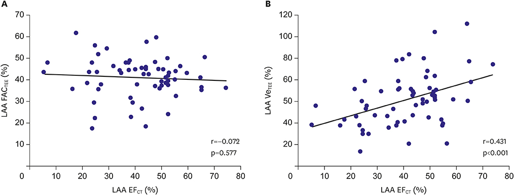
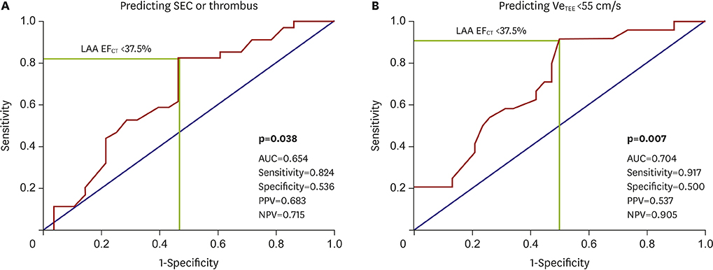
SEC or thrombus was observed in 45.2%. EFCT and VeTEE were significantly correlated (r=0.452, p < 0.001). However, fractional area change measured by TEE showed no correlation with VeTEE (r=0.085, p=0.512). EFCT < 37.5% best predicted SEC or thrombus in the patients with valve disease who underwent 4DCT and TEE (area under the curve, 0.654; p=0.038).
CONCLUSIONS
In the patients who underwent 4DCT for cardiac valve evaluation before surgery, EFCT by volume analysis might have additional role to evaluate LAA function and estimate the risk of thrombus.
Keyword
MeSH Terms
Figure
Cited by 1 articles
-
Computed Tomography for Assessment of Left Atrial Appendage Function
Jin Joo Park
Korean Circ J. 2019;49(2):181-182. doi: 10.4070/kcj.2018.0355.
Reference
-
1. Handke M, Harloff A, Hetzel A, Olschewski M, Bode C, Geibel A. Left atrial appendage flow velocity as a quantitative surrogate parameter for thromboembolic risk: determinants and relationship to spontaneous echocontrast and thrombus formation--a transesophageal echocardiographic study in 500 patients with cerebral ischemia. J Am Soc Echocardiogr. 2005; 18:1366–1372.
Article2. Delgado V, Tops LF, Schuijf JD, et al. Assessment of mitral valve anatomy and geometry with multislice computed tomography. JACC Cardiovasc Imaging. 2009; 2:556–565.
Article3. Jilaihawi H, Kashif M, Fontana G, et al. Cross-sectional computed tomographic assessment improves accuracy of aortic annular sizing for transcatheter aortic valve replacement and reduces the incidence of paravalvular aortic regurgitation. J Am Coll Cardiol. 2012; 59:1275–1286.
Article4. Messé SR, Acker MA, Kasner SE, et al. Stroke after aortic valve surgery: results from a prospective cohort. Circulation. 2014; 129:2253–2261.5. Russo A, Grigioni F, Avierinos JF, et al. Thromboembolic complications after surgical correction of mitral regurgitation incidence, predictors, and clinical implications. J Am Coll Cardiol. 2008; 51:1203–1211.6. Hahn RT, Abraham T, Adams MS, et al. Guidelines for performing a comprehensive transesophageal echocardiographic examination: recommendations from the American society of echocardiography and the society of cardiovascular anesthesiologists. J Am Soc Echocardiogr. 2013; 26:921–964.
Article7. Fatkin D, Kelly RP, Feneley MP. Relations between left atrial appendage blood flow velocity, spontaneous echocardiographic contrast and thromboembolic risk in vivo. J Am Coll Cardiol. 1994; 23:961–969.
Article8. Romero J, Husain SA, Kelesidis I, Sanz J, Medina HM, Garcia MJ. Detection of left atrial appendage thrombus by cardiac computed tomography in patients with atrial fibrillation: a meta-analysis. Circ Cardiovasc Imaging. 2013; 6:185–194.9. Palmer S, Child N, de Belder MA, Muir DF, Williams P. Left atrial appendage thrombus in transcatheter aortic valve replacement: incidence, clinical impact, and the role of cardiac computed tomography. JACC Cardiovasc Interv. 2017; 10:176–184.10. Iwasaki K, Matsumoto T, Kawada S. Potential utility of multidetector computed tomography to identify both cardiac embolic sources and coronary artery disease in patients with embolic stroke. Cardiology. 2016; 133:205–210.
Article11. Hur J, Kim YJ, Lee HJ, et al. Cardiac computed tomographic angiography for detection of cardiac sources of embolism in stroke patients. Stroke. 2009; 40:2073–2078.
Article12. Ko SB, Choi SI, Chun EJ, et al. Role of cardiac multidetector computed tomography in acute ischemic stroke: a preliminary report. Cerebrovasc Dis. 2010; 29:313–320.
Article13. Kim IC, Chang S, Hong GR, et al. Comparison of cardiac computed tomography with transesophageal echocardiography for identifying vegetation and intracardiac complications in patients with infective endocarditis in the era of 3-dimensional images. Circ Cardiovasc Imaging. 2018; 11:e006986.
Article14. Takahashi K, Stanford W. Multidetector CT of the thoracic aorta. Int J Cardiovasc Imaging. 2005; 21:141–153.
Article15. Yoo J, Song D, Baek JH, et al. Poor outcome of stroke patients with atrial fibrillation in the presence of coexisting spontaneous echo contrast. Stroke. 2016; 47:1920–1922.
Article16. Zhao Y, Ji L, Liu J, et al. Intensity of left atrial spontaneous echo contrast as a correlate for stroke risk stratification in patients with nonvalvular atrial fibrillation. Sci Rep. 2016; 6:27650.
Article17. Daniel WG, Nellessen U, Schröder E, et al. Left atrial spontaneous echo contrast in mitral valve disease: an indicator for an increased thromboembolic risk. J Am Coll Cardiol. 1988; 11:1204–1211.
Article18. Kupczyńska K, Kasprzak JD, Michalski B, Lipiec P. Prognostic significance of spontaneous echocardiographic contrast detected by transthoracic and transesophageal echocardiography in the era of harmonic imaging. Arch Med Sci. 2013; 9:808–814.
Article
- Full Text Links
- Actions
-
Cited
- CITED
-
- Close
- Share
- Similar articles
-
- Comparisons of the Left Ventricular Ejection Fraction Between M-Mode, Two-Dimension Echocardiographic Studies and Digital Cardiac Imaging(DCI) System in Ventricular Septal Defects*
- Left Atrial Thrombus and Spontaneous Echo Contrast in Mitral Valvular Heart Disease : Its Clinical Significance and the Role of Transesophageal Echocardiography
- Cardioembolic Stroke in Atrial Fibrillation-Rationale for Preventive Closure of the Left Atrial Appendage
- Relation between Left Atrial Size and Atrial Fibrillation
- Assessment of Left Atrial Appendage Flow Pattern Using Multiplane Transesophageal Echocardiography in Patients with Nonrheumatic Atrial Fibrillation and Ischemic Stroke


