J Korean Ophthalmol Soc.
2019 Jan;60(1):96-101. 10.3341/jkos.2019.60.1.96.
A Case of Leber Hereditary Optic Neuropathy Showing Optic Disc Hyperfluorescence
- Affiliations
-
- 1Department of Ophthalmology, Asan Medical Center, University of Ulsan College of Medicine, Seoul, Korea. htlim@amc.seoul.kr
- KMID: 2431847
- DOI: http://doi.org/10.3341/jkos.2019.60.1.96
Abstract
- PURPOSE
We report an unusual case of Leber hereditary optic neuropathy presenting with optic disc hyperfluorescence.
CASE SUMMARY
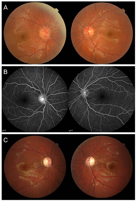
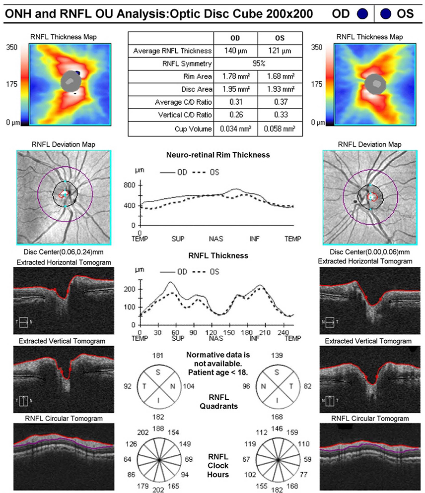
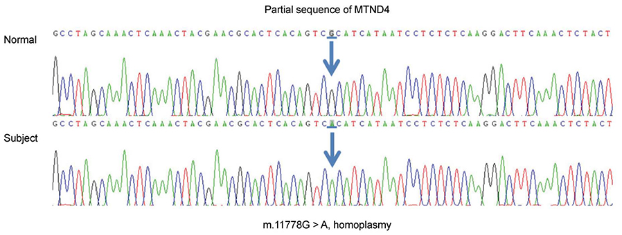
A 17-year-old male with sequential painless visual loss 3 weeks apart affecting first the left and then the right eye presented to our neuro-ophthalmology clinic. His best-corrected visual acuity was counting fingers in the right eye and 0.32 in the left eye. Fundus examination showed mild optic disc edema and hyperemia in both eyes, which were worse in the right eye. Fluorescein angiography revealed dye leakage from the right optic disc in the late phase. The results of magnetic resonance imaging of the brain and spinal cord were normal, and lumbar puncture study was unremarkable. Mitochondrial DNA sequencing revealed a pathognomonic 11778 mutation for Leber hereditary optic neuropathy. His vision deteriorated to 0.03 in both eyes 6 months later, but slowly started to improve 11 months after onset. At 2 years, his corrected visual acuity was 0.2 in both eyes.
CONCLUSIONS
To our knowledge, this is the first report of optic disc hyperfluorescence in Leber hereditary optic neuropathy. This finding suggests that this mitochondrial optic neuropathy can masquerade as optic neuritis.
MeSH Terms
Figure
Reference
-
1. Newman NJ. Hereditary optic neuropathies: from the mitochondria to the optic nerve. Am J Ophthalmol. 2005; 140:517–523.
Article2. Yu-Wai-Man P, Griffiths PG, Brown DT, et al. The epidemiology of Leber hereditary optic neuropathy in the North East of England. Am J Hum Genet. 2003; 72:333–339.3. Wallace DC, Singh G, Lott MT, et al. Mitochondrial DNA mutation associated with Leber's hereditary optic neuropathy. Science. 1988; 242:1427.
Article4. Newman NJ, Lott MT, Wallace DC. The clinical characteristics of pedigrees of Leber's hereditary optic neuropathy with the 11778 mutation. Am J Ophthalmol. 1991; 111:750–762.
Article5. Smith J, Hoyt WF, Susac JO. Ocular fundus in acute leber optic neuropathy. Arch Ophthalmol. 1973; 90:349–354.
Article6. Riordan-Eva P, Harding AE. Leber's hereditary optic neuropathy: the clinical relevance of different mitochondrial DNA mutations. J Med Genet. 1995; 32:81–87.
Article7. Sadun AA, Salomao SR, Berezovsky A, et al. Subclinical carriers and conversions in Leber hereditary optic neuropathy: a prospective psychophysical study. Trans Am Ophthalmol Soc. 2006; 104:51–61.8. Hsu TK, Wang AG, Yen MY, Liu JH. Leber's hereditary optic neuropathy masquerading as optic neuritis with spontaneous visual recovery. Clin Exp Optom. 2014; 97:84–86.
Article9. Schachat AP, Wilkinson CP, Hinton DR, et al. Ryan's Retina. 6th ed. 1. Toronto: Elsevier;2017. p. 19–44.10. Sadun AA, Win PH, Ross-Cisneros FN, et al. Leber's hereditary optic neuropathy differentially affects smaller axons in the optic nerve. Trans Am Ophthalmol Soc. 2000; 98:223–232. discussion 232-5.11. Nikoskelainen EK, Huoponen K, Juvonen V, et al. Ophthalmologic findings in Leber hereditary optic neuropathy, with special reference to mtDNA mutations. Ophthalmology. 1996; 103:504–514.
Article12. Piotrowska A, Korwin M, Bartnik E, Tońska K. Leber hereditary optic neuropathy-historical report in comparison with the current knowledge. Gene. 2015; 555:41–49.13. Wan MJ, Adebona O, Benson LA, et al. Visual outcomes in pediatric optic neuritis. Am J Ophthalmol. 2014; 158:503–507.e2.
Article14. Golnik K. Nonglaucomatous optic atrophy. Neurol Clin. 2010; 28:631–640.
Article
- Full Text Links
- Actions
-
Cited
- CITED
-
- Close
- Share
- Similar articles
-
- Clinical Manifestations of Leber's Hereditary Optic Neuropathy with 11778 mtDNA Mutation
- Leber's Hereditary Optic Neuropathy in Two Brothers of a Family
- A Mitochondrial Mutation in Leber's Hereditary Optic Neuropathy
- Three Cases of Leber's Hereditary Optic Atrophy in One Family
- A Case of Leber's Hereditary Optic Nouropathy Showing 11778 Point Mutation of Mitochondrial DNA


