Magnetization Transfer Contrast Angiography for Organized Thrombosed Intracranial Aneurysm in TOF MR Angiography: a Case Report
- Affiliations
-
- 1Department of Neurosurgery, Kyungpook National University, School of Medicine, Daegu, Korea.
- 2Department of Radiology, Kyungpook National University, School of Medicine, Daegu, Korea. leehuijoong@knu.ac.kr
- KMID: 2431113
- DOI: http://doi.org/10.13104/imri.2018.22.4.266
Abstract
- A 66-year-old woman was referred for treatment of incidental detection of two intracranial aneurysms. Time-of-flight MR angiography (TOF MRA) revealed two aneurysms at the M1 segment of the right middle cerebral artery, and clinoid segment of left internal carotid artery, respectively. On digital subtraction angiography, there was a saccular aneurysm on the left internal carotid artery, but the other aneurysm was not detected on the right middle cerebral artery. Based on comprehensive review of imaging findings, organized thrombosed aneurysm was judged as the most likely diagnosis. In the presented report, magnetization transfer (MT) pulse to TOF MRA was used, to differentiate aneurysm-mimicking lesion on TOF MRA. We report that MT technique could be effective in differentiating true aneurysm, from possible T1 high signal artifact on TOF MRA.
Keyword
MeSH Terms
Figure
-
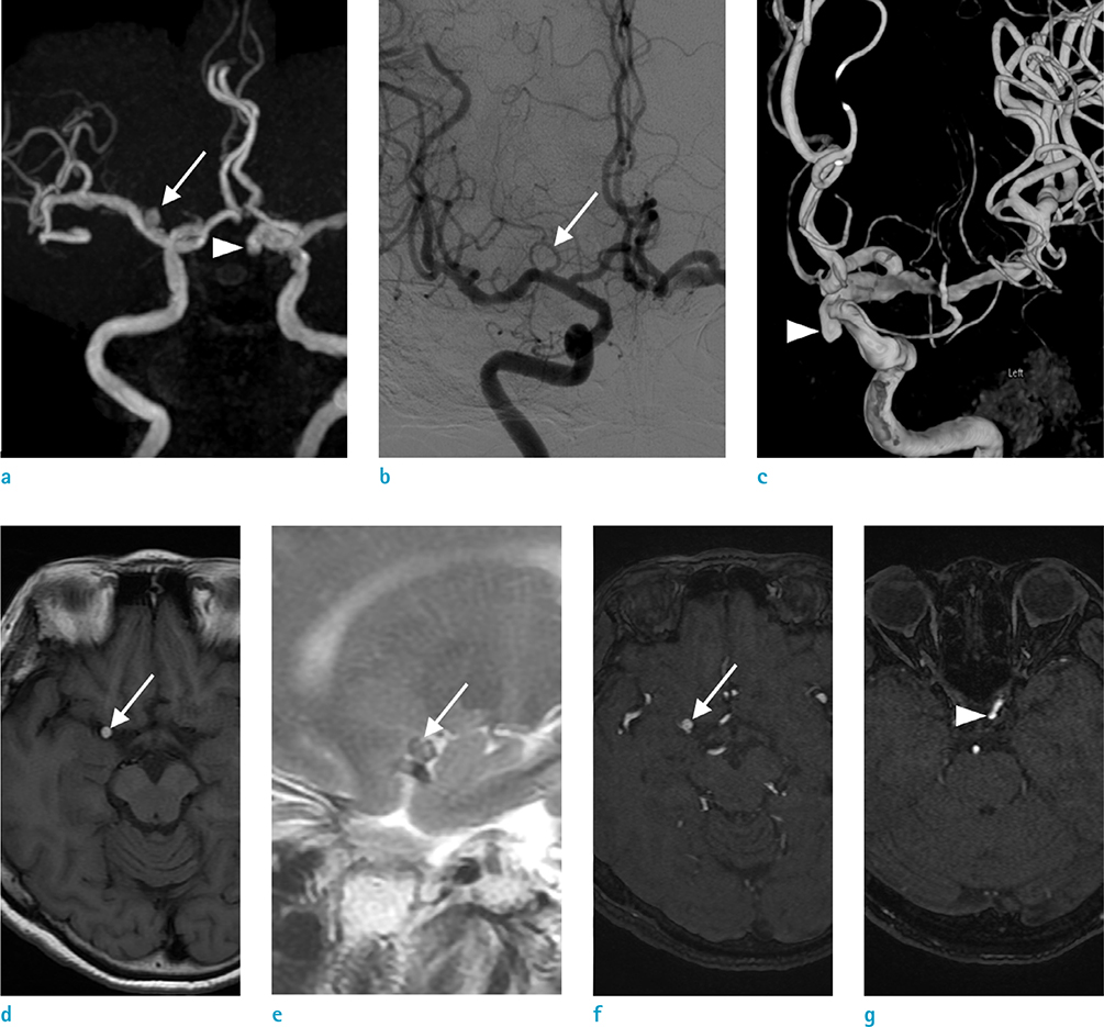
Fig. 1 66-year-old woman with a diagnostic pitfall on TOF MR angiography. TOF MRA (a) shows two intracranial aneurysms; one is located at the M1 segment of right middle cerebral artery (MCA; arrow) and the clinoid segment of left internal carotid artery (ICA; arrowhead). Aneurysm was invisible (arrow) digital subtraction angiography (b). In contrast, saccular aneurysm from the left ICA is noted on 3D Rotational angiography (c, arrowhead). The lesion at the vicinity of the right MCA shows high signal intensity (arrow) on T1 (d) and low signal intensity (arrow) on T2 weight MR image (e). High signal intensity lesions suggestive aneurysms (arrow) at the vicinity of right MCA (f) and ophthalmic segment of left ICA (arrowhead) (g) are noted on the source images of TOF MRA.
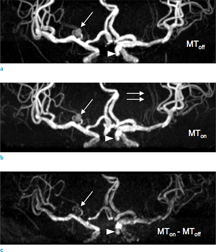
Fig. 2 Magnetization transfer contrast angiography. As compared with an aneurysm (arrow) on MToff (a), better conspicuity of lenticulostriate arteries (double arrows) is noted on MTon angiography (b). MTC subtraction MRA (c) as a result of subtraction of MTon (b) from MToff (a) shows no aneurysm (arrow) in the right middle cerebral artery, contrary to well definition of aneurysm (arrowhead) at right ICA.
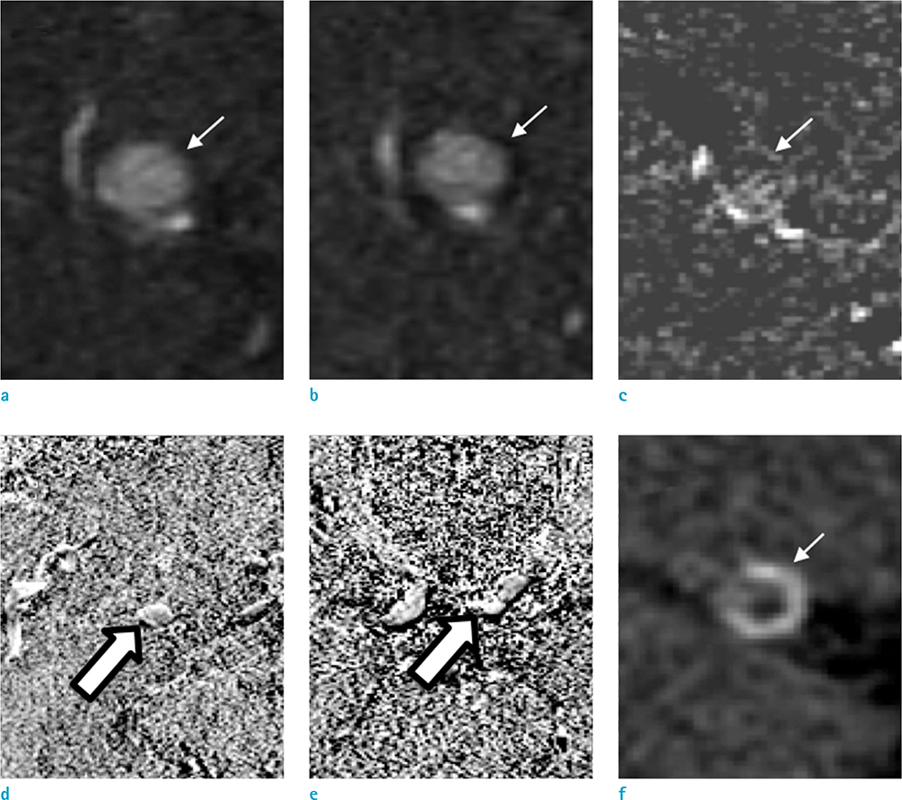
Fig. 3 Phase shift TOF MRA (a: in-phase, b: out-of-phase) did not showed chemical artifact around small hyperintense lesion (arrows) in the right middle cerebral artery. Source image of magnetization transfer contrast MRA (c) shows low signal intensity of pitfall (arrow). MTR of pitfall (open arrow) (d) and true lesion (open arrow) (e) was 0.170 ± 0.230, and 0.421 ± 0.209, respectively. CT (f) shows high-density wall (Hounsfield unit = 134.2 ± 30.2) with relatively low-density core (arrow) (Hounsfield unit = 64.4 ± 20.2) on the center of lesion.
Reference
-
1. Wolff SD, Balaban RS. Magnetization transfer contrast (MTC) and tissue water proton relaxation in vivo. Magn Reson Med. 1989; 10:135–144.2. Henkelman RM, Stanisz GJ, Graham SJ. Magnetization transfer in MRI: a review. NMR Biomed. 2001; 14:57–64.
Article3. Pike GB, Hu BS, Glover GH, Enzmann DR. Magnetization transfer time-of-flight magnetic resonance angiography. Magn Reson Med. 1992; 25:372–379.
Article4. Edelman RR, Ahn SS, Chien D, et al. Improved time-of-flight MR angiography of the brain with magnetization transfer contrast. Radiology. 1992; 184:395–399.
Article5. Atkinson D, Brant-Zawadzki M, Gillan G, Purdy D, Laub G. Improved MR angiography: magnetization transfer suppression with variable flip angle excitation and increased resolution. Radiology. 1994; 190:890–894.
Article6. Nezafat R, Han Y, Peters DC, et al. Coronary magnetic resonance vein imaging: imaging contrast, sequence, and timing. Magn Reson Med. 2007; 58:1196–1206.
Article7. Lin W, Tkach JA, Haacke EM, Masaryk TJ. Intracranial MR angiography: application of magnetization transfer contrast and fat saturation to short gradient-echo, velocity-compensated sequences. Radiology. 1993; 186:753–761.
Article8. Anderson CM, Saloner D, Tsuruda JS, Shapeero LG, Lee RE. Artifacts in maximum-intensity-projection display of MR angiograms. AJR Am J Roentgenol. 1990; 154:623–629.
Article9. Wilcock DJ, Jaspan T, Worthington BS. Problems and pitfalls of 3-D TOF magnetic resonance angiography of the intracranial circulation. Clin Radiol. 1995; 50:526–532.
Article10. Wermer MJ, van der Schaaf IC, Velthuis BK, Majoie CB, Albrecht KW, Rinkel GJ. Yield of short-term follow-up CT/MR angiography for small aneurysms detected at screening. Stroke. 2006; 37:414–418.
Article11. Soila KP, Viamonte M Jr, Starewicz PM. Chemical shift misregistration effect in magnetic resonance imaging. Radiology. 1984; 153:819–820.
Article12. Sankhla SK, Gunawardena WJ, Coutinho CM, Jones AP, Keogh AJ. Magnetic resonance angiography in the management of aneurysmal subarachnoid haemorrhage: a study of 51 cases. Neuroradiology. 1996; 38:724–729.
Article13. Litt AW. MR angiography of intracranial aneurysms: proceed, but with caution. AJNR Am J Neuroradiol. 1994; 15:1615–1616.14. Wardlaw JM, White PM. The detection and management of unruptured intracranial aneurysms. Brain. 2000; 123(Pt 2):205–221.
Article15. Okahara M, Kiyosue H, Yamashita M, et al. Diagnostic accuracy of magnetic resonance angiography for cerebral aneurysms in correlation with 3D-digital subtraction angiographic images: a study of 133 aneurysms. Stroke. 2002; 33:1803–1808.16. Horikoshi T, Fukamachi A, Nishi H, Fukasawa I. Detection of intracranial aneurysms by three-dimensional time-of-flight magnetic resonance angiography. Neuroradiology. 1994; 36:203–207.
Article17. Adams WM, Laitt RD, Jackson A. The role of MR angiography in the pretreatment assessment of intracranial aneurysms: a comparative study. AJNR Am J Neuroradiol. 2000; 21:1618–1628.18. Kemmling A, Noelte I, Gerigk L, Singer S, Groden C, Scharf J. A diagnostic pitfall for intracranial aneurysms in time-of-flight MR angiography: small intracranial lipomas. AJR Am J Roentgenol. 2008; 190:W62–W67.
Article19. Trungu S, Bruzzaniti P, Forcato S, Cimatti M, Raco A. Completely thrombosed distal middle cerebral artery aneurysm mimicking a cavernous angioma: case report and review of the literature. World Neurosurg. 2017; 103:955.
Article20. Qiao Y, Hallock KJ, Hamilton JA. Magnetization transfer magnetic resonance of human atherosclerotic plaques ex vivo detects areas of high protein density. J Cardiovasc Magn Reson. 2011; 13:73.
Article
- Full Text Links
-
- Actions
-
Cited
- CITED
-
- Close
- Share
-
- Similar articles
-
- Imaging Features of Intracranial Calcified Aneurysm: Report of 4 Cases
- Completely Thrombosed Contralateral Internal Carotid Artery Aneurysm Combined with Ruptured Anterior Communicating Artery Aneurysm: Case Report
- Comparison of MR Angiography with Conventional Angiography in Cervical and Intracranial Vascular Diseases
- Comparison of Magnetic Resonance Angiography and CT Angiography in the Evaluation of Intracranial Aneurysm
- Rupture of Giant Posterior Communicating Artery Aneurysm During Carotid Angiography: A Case Report
