Cytohesin-2 Is Upregulated in Malignant Melanoma and Contributes to Tumor Growth
- Affiliations
-
- 1Molecular Cancer Research, Soonchunhyang University College of Medicine, Cheonan, Korea.
- 2Department of Dermatology, Soonchunhyang University Seoul Hospital, Seoul, Korea. mkcho@schmc.ac.kr
- 3Department of Plastic and Reconstructive Surgery, Soonchunhyang University Cheonan Hospital, Cheonan, Korea.
- 4Department of Dermatology, Soonchunhyang University Bucheon Hospital, Bucheon, Korea.
- KMID: 2430811
- DOI: http://doi.org/10.5021/ad.2019.31.1.93
Abstract
- No abstract available.
MeSH Terms
Figure
-
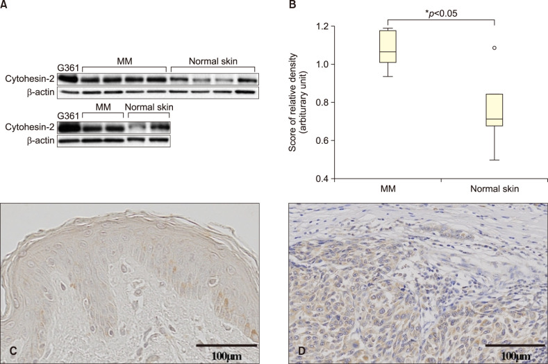
Fig. 1 Western blot analysis of Cytohesin-2 in malignant melanoma. (A) Cytohesin-2 was expressed in malignant melanoma (MM) tissues, but was decreased in normal human skin tissues. (B) The relative protein expression of Cytohesin-2 according to the Mann-Whitney U-test was analyzed. Representative immunohistochemistry (IHC) staining for Cytohesin-2 protein expression in paraffin-embedded normal skin tissue and MM. Brown color spots were positively stained cells. (C) Negative staining of Cytohesin-2 in normal skin (IHC stain, ×400). (D) Mild to moderate staining of Cytohesin-2 in MM (IHC stain, ×400). β-actin, beta-actin.
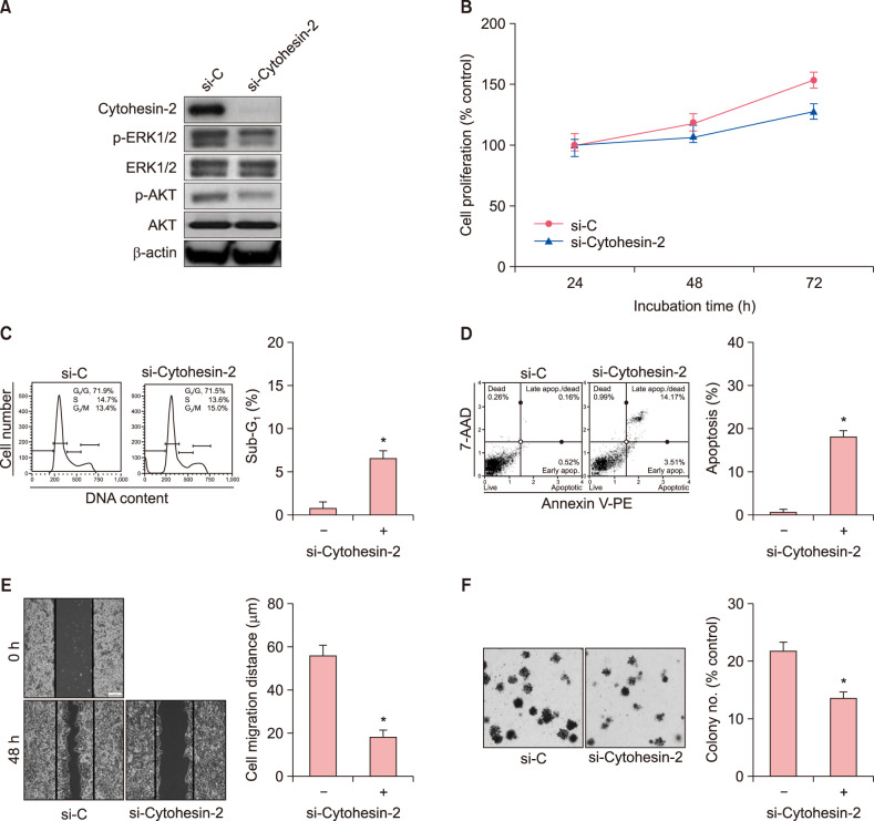
Fig. 2 G361 cells were transfected with Cytohesin-2-specific siRNA. (A) Western blot of Cytohesin-2, p-ERK1/2, ERK, p-AKT and AKT protein. (B) The percentage of viable cells was measured by MTT assay. (C) Cell distribution in G0/G1, S, and G2/M phases was analyzed using flow cytometry after staining with propidium iodide (20 µg/ml). (D) The percentage of apoptotic cells after Annexin V-PE binding was analyzed using a Muse Cell Analyzer. (E) Representative images of the wound healing assay in G361 cells. (F) Colony formation assay for G361 cells transfected with control and si-Cytohesin-2. Error bars represent mean±standard deviation for three independent experiments. *p<0.05 compared to respective controls. β-actin: beta-actin, si-C: StealthTM RNAi control, si-Cytohesin-2: small-interfering Cytohesin-2, p-: phospho-, ERK: signal-regulated protein kinases, 7-AAD: 7-aminoactinomycin D.
Reference
-
1. Moss J, Vaughan M. Molecules in the ARF orbit. J Biol Chem. 1998; 273:21431–21434. PMID: 9705267.
Article2. Kolanus W. Guanine nucleotide exchange factors of the cytohesin family and their roles in signal transduction. Immunol Rev. 2007; 218:102–113. PMID: 17624947.
Article3. Casanova JE. Regulation of Arf activation: the Sec7 family of guanine nucleotide exchange factors. Traffic. 2007; 8:1476–1485. PMID: 17850229.
Article4. Lim J, Zhou M, Veenstra TD, Morrison DK. The CNK1 scaffold binds cytohesins and promotes insulin pathway signaling. Genes Dev. 2010; 24:1496–1506. PMID: 20634316.
Article5. Pan T, Sun J, Hu J, Hu Y, Zhou J, Chen Z, et al. Cytohesins/ARNO: the function in colorectal cancer cells. PLoS One. 2014; 9:e90997. PMID: 24618737.
Article6. Xu K, Gao J, Yang X, Yao Y, Liu Q. Cytohesin-2 as a novel prognostic marker for hepatocellular carcinoma. Oncol Rep. 2013; 29:2211–2218. PMID: 23545718.
Article7. Santy LC, Casanova JE. Activation of ARF6 by ARNO stimulates epithelial cell migration through downstream activation of both Rac1 and phospholipase D. J Cell Biol. 2001; 154:599–610. PMID: 11481345.
Article8. Muralidharan-Chari V, Hoover H, Clancy J, Schweitzer J, Suckow MA, Schroeder V, et al. ADP-ribosylation factor 6 regulates tumorigenic and invasive properties in vivo. Cancer Res. 2009; 69:2201–2209. PMID: 19276388.
- Full Text Links
-
- Actions
-
Cited
- CITED
-
- Close
- Share
-
- Similar articles
-
- Malignant Melanoma on Congenital Melanocytic Nevus
- A Case of Conjunctival Malignant Melanoma with Extensive Corneal Displacement
- A Case of Nodular Melanoma with a Family History of Difficult Clinical Diagnosis
- Three Cases of Malignant Melanoma Possibly Arising in a Long Standing Melanocytic Nevus
- A Case of Primary Malignant Leptomeningeal Melanoma of the Spinal Cord: Case Report
