Ann Dermatol.
2019 Feb;31(1):66-69. 10.5021/ad.2019.31.1.66.
Isolated Plantar Vein Thrombosis Resembling a Corn with a Bruise
- Affiliations
-
- 1Department of Dermatology, Kangdong Sacred Heart Hospital, College of Medicine, Hallym University, Seoul, Korea. hje150273@naver.com
- KMID: 2430803
- DOI: http://doi.org/10.5021/ad.2019.31.1.66
Abstract
- Plantar vein thrombosis, rarely-reported disease, is usually accompanied by pain and tenderness in the plantar region and should be differentiated from other dermatological conditions causing plantar pain, such as hemorrhagic corn/callus, plantar epidermal cyst, verruca, or plantar fibromatosis. A 52-year-old man presented with a violaceous tender subcutaneous nodule overlying a hyperkeratotic plaque on his sole. Initially, he thought it was a corn and applied keratolytic agents, which failed to work. Sonography revealed a well-demarcated mass with increased peripheral vascularity. His pain was relieved after a complete wide excision, which confirmed the mass to be plantar vein thrombosis after histopathological examination.
Keyword
MeSH Terms
Figure
-
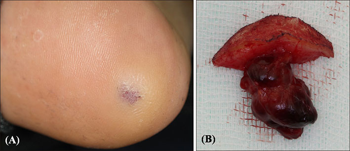
Fig. 1 (A) A bruise-like violaceous nodule 2.5 cm in diameter, on the heel side of the right sole. (B) A dark reddish rubbery, lobulated nodule in the subcutaneous layer.
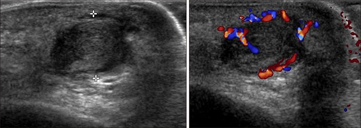
Fig. 2 A 1.9×1.2 cm sized, well-marginated, lobulated, heterogeneous hypoechoic mass surrounded by a thin anechoic area with increased peripheral vascularity.
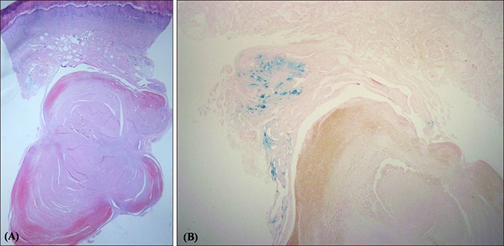
Fig. 3 (A) Several variable thick and plicated walled small muscular vessels in the dermis and its abutting thrombus consisting of aggregated erythrocytes and eosinophilic fibrin in the lumen of a dilated vein in the subcutaneous layer (H&E, ×10). (B) Granular bluish deposit (hemosiderin) within and around mural walls of muscular vessels (Iron stain, ×20).
Reference
-
1. Wallace GF. Dermatologic causes of heel pain. Clin Podiatr Med Surg. 2010; 27:407–416.
Article2. Bernathova M, Bein E, Bendix N, Bodner G. Sonographic diagnosis of plantar vein thrombosis: report of 3 cases. J Ultrasound Med. 2005; 24:101–103.3. Siegal DS, Wu JS, Brennan DD, Challies T, Hochman MG. Plantar vein thrombosis: a rare cause of plantar foot pain. Skeletal Radiol. 2008; 37:267–269.
Article4. Long A, Bura-Riviere A, Sapoval M. [Plantar venous thrombosis and anticardiolipin antibody syndrome. Case report]. J Mal Vasc. 2004; 29:39–40. French.5. Cavezzi A. Isolated thrombosis of plantar veins. Case report. Minerva Cardioangiol. 1999; 47:309–313.6. Legrand MS, Papon X, Leftheriotis G, Saumet JL. [Isolated plantar venous thrombosis. Report of a case]. J Mal Vasc. 1997; 22:364–365. French.7. Lee KM, Park JH, Min KH, Kim EK. Epidermal cyst on the sole. Arch Plast Surg. 2013; 40:475–476.
Article8. Bae JM, Kang H, Kim HO, Park YM. Differential diagnosis of plantar wart from corn, callus and healed wart with the aid of dermoscopy. Br J Dermatol. 2009; 160:220–222.
Article9. Thomas VD, Snavely NR, Lee KK, Swanson NA. Benign epithelial tumors, hamartomas, and hyperplasias. In : Goldsmith LA, Katz SI, Gilchrest BA, Paller AS, Leffell DJ, Wolff K, editors. Fitzpatrick's dermatology in general medicine. 8th ed. New York: McGraw-Hill;2012. p. 1319–1336.10. Robbin MR, Murphey MD, Temple HT, Kransdorf MJ, Choi JJ. Imaging of musculoskeletal fibromatosis. Radiographics. 2001; 21:585–600.
Article11. Geiger C, Rademacher A, Chappell D, Sadeghi-Azandaryani M, Heyn J. Plantar vein thrombosis due to busy night duty on intensive care unit. Clin Appl Thromb Hemost. 2011; 17:232–234.
Article12. Fabro M, Fabro SR, Sales RS, Machado CA, de Araújo GL. Plantar vein thrombosis: a rare differential diagnosis in patients with plantar pain. Radiol Bras. 2015; 48:399–400.
Article13. Houdek MT, Warneke JA, Pollard CM, Lindgren EA, Taljanovic MS. Giant epidermal cyst of the gluteal region. Radiol Case Rep. 2015; 5:476.
Article
- Full Text Links
-
- Actions
-
Cited
- CITED
-
- Close
- Share
-
- Similar articles
-
- A Case of Isolated Plantar Digital Vein Thrombosis
- A Case of Isolated Plantar Vein Thrombosis
- Plantar Corn Caused by Epidermal Cyst
- Isolated Splenic Vein Thrombosis Associated with Acute Pancreatitis
- Concurrent Isolated Cortical Vein Thrombosis and Pulmonary Thromboembolism as An Initial Presentation of Protein S Deficiency
