Massive splenomegaly: flow cytometry as a diagnostic tool for systemic mastocytosis
- Affiliations
-
- 1Department of Hematology, Sir Ganga Ram Hospital, New Delhi, India. drjasmita@gmail.com
- 2Department of Clinical Hematology, Sir Ganga Ram Hospital, New Delhi, India.
- KMID: 2429327
- DOI: http://doi.org/10.5045/br.2018.53.3.251
Abstract
- No abstract available.
Figure
-
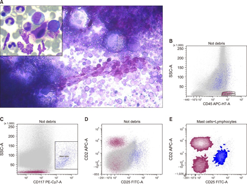
Fig. 1 (A) Toluidine blue staining shows mast cells in a marrow particle (toluidine blue, ×200). The inset shows spindle cell morphology of mast cells (H&E, ×400). (B) CD45 vs side scatter plot (SSC) to gate lymphocytes (reddish brown). (C) Gating for mast cells was done on CD117 vs SSC dot plot. Bright CD117+ cells are gated as mast cells (dark blue). (D, E) Dot plots and color density plots respectively showing an aberrant expression of CD2 and CD25 on the mast cell population. The intensity of CD2 is lesser than the normal T lymphocytes.
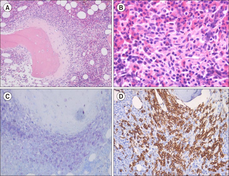
Fig. 2 (A) Paratrabecular mast cell aggregate showing new bone formation (H&E, ×100). (B) A high-power view shows the atypical morphology of mast cells with a predominance of oval- and spindle-shaped mast cells. Eosinophils are interspersed in the aggregates (H&E, ×400). (C) Toluidine blue staining shows metachromasia in mast cell aggregates (×200). (D) CD117 immunohistochemistry on bone marrow biopsy is positive in paratrabecular mast cell aggregates (×200).
Reference
-
1. Arber DA, Orazi A, Hasserjian R, et al. The 2016 revision to the World Health Organization classification of myeloid neoplasms and acute leukemia. Blood. 2016; 127:2391–2405. PMID: 27069254.
Article2. Horny HP, Metcalfe DD, Bennett JM, et al. Mastocytosis. In : Swerdlow SH, Campo E, Harris NL, editors. WHO classification of tumours of haematopoietic and lymphoid tissues. 4th ed. Lyon, France: IARC Press;2008. p. 54–63.3. Arredondo AR, Gotlib J, Shier L, et al. Myelomastocytic leukemia versus mast cell leukemia versus systemic mastocytosis associated with acute myeloid leukemia: a diagnostic challenge. Am J Hematol. 2010; 85:600–606. PMID: 20658589.
Article4. Pozdnyakova O, Kondtratiev S, Li B, Charest K, Dorfman DM. High-sensitivity flow cytometric analysis for the evaluation of systemic mastocytosis including the identification of a new flow cytometric criterion for bone marrow involvement. Am J Clin Pathol. 2012; 138:416–424. PMID: 22912359.
Article
- Full Text Links
-
- Actions
-
Cited
- CITED
-
- Close
- Share
-
- Similar articles
-
- Analysis of T Cells Using Flow Cytometry
- A Case of Adult-onset Urticaria Pigmentosa with Bone Involvement
- Solitary mastocytoma presenting at birth
- The Value of Urine Cytology and Flow Cytometry in Superficial Bladder Tumors
- A case of lymphocytic variant hypereosinophilic syndrome with sub-diagnostic systemic mastocytosis
