First Case of Necrotizing Fasciitis Caused by Skermanella aerolata Infection Mimicking Vibrio Sepsis
- Affiliations
-
- 1Department of Infectious Disease, Jeju National University School of Medicine, Jeju, Korea.
- 2Department of Internal Medicine, School of Medicine, Kyungpook National University, Daegu, Korea.
- 3Department of Molecular Cell Biology and Samsung Medical Center, Sungkyunkwan University School of Medicine, Suwon, Korea. ksko@skku.edu
- 4Department of Microbiology and Immunology, Jeju National University School of Medicine, Jeju, Korea.
- KMID: 2429128
- DOI: http://doi.org/10.3343/alm.2018.38.6.604
Abstract
- No abstract available.
MeSH Terms
Figure
-
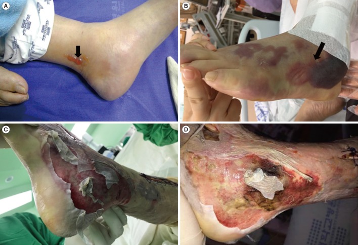
Fig. 1 Development of infected area. Swelling and small vesicles (A, black arrow) detected on the left ankle area of the patient on presentation to the emergency department. The lesions changed to hemorrhagic bullae (B, black arrow) extending from the foot to shin area within about 12 hours after admission. In the operating room, the skin of the left foot detached easily along the lateral aspect (C) with progression to an ischemic color change (D) along the medial aspect.
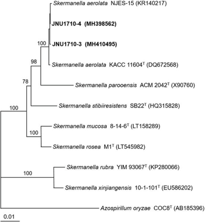
Fig. 2 Phylogenetic tree of isolates JNU1710-3 (from tissue) and JNU1710-4 (from serum) and species of Skermanella based on 16S rRNA gene sequences. The tree was reconstructed by the neighbor-joining method (MEGA 5.10), and Azospirillium oryzae COC8T was used as an outgroup. Numbers on branching nodes are percentages of 1,000 bootstrap replications; only values ≥ 50% are shown. The scale bar represents one substitution per 100 nucleotides.
Reference
-
1. Sly LI, Stackebrandt E. Description of Skermanella parooensis gen. nov., sp. nov. to accommodate Conglomeromonas largomobilis subsp. parooensis following the transfer of Conglomeromonas largomobilis subsp. largomobilis to the genus Azospirillum. Int J Syst Bacteriol. 1999; 49:541–544.2. Subhash Y, Yoon DE, Lee SS. Skermanella mucosa sp. nov., isolated from crude oil contaminated soil. Antonie van Leeuwenhoek. 2017; 110:1053–1060. PMID: 28501914.3. Kalwasińska A, Felföldi T, Szabó A, Deja-Sikora E, Kosobucki K, Walczak M. Microbial communities associated with the anthropogenic highly alkaline environment of a saline soda lime, Poland. Antonie van Leeuwenhoek. 2017; 110:945–962. PMID: 28382378.4. Park JY, Jeon S, Kim JY, Park M, Kim S. Multiplex real-time polymerase chain reaction assays for simultaneous detection of Vibrio cholera, Vibrio parahaemolyticus, and Vibrio vulnificus. Osong Public Health Res Perspect. 2013; 4:133–139. PMID: 24159544.5. Al Masalma M, Armougom F, Scheld WM, Dufour H, Roche PH, Drancourt M, et al. The expansion of the microbiological spectrum of brain abscess with use of multiple 16S ribosomal DNA sequencing. Clin Infect Dis. 2009; 48:1169–1178. PMID: 19335164.6. Yoon SH, Ha SM, Kwon S, Lim J, Seo H, Chun J. Introducing EzBioCloud: a taxonomically united database of 16S rRNA and whole genome assemblies. Int J Syst Evol Microbiol. 2017; 67:1613–1617. PMID: 28005526.7. Weon HY, Kim BY, Hong SB, Joa JH, Nam SS, Lee KH, et al. Skermanella aerolata sp. nov., isolated from air, and emended description of the genus Skermanella. Int J Syst Evol Microbiol. 2007; 57:1539–1542. PMID: 17625190.8. Wang L, Li J, Yang F, Yaoyao E, Raza W, Huang Q, et al. Application of bioorganic fertilizer significantly increased apple yields and shaped bacterial community structure in orchard soil. Microb Ecol. 2017; 73:404–416. PMID: 27670433.9. Horseman MA, Surani S. A comprehensive review of Vibrio vulnificus: an important cause of severe sepsis and skin and soft-tissue infection. Int J Infect Dis. 2011; 15:e157–e166. PMID: 21177133.10. Lee KH, Heo ST, Kim YR, Pang IC. Isolation of Vibrio vulnificus from seawater and emerging Vibrio vulnificus septicemia on Jeju Island. Infect Chemother. 2014; 46:106–109. PMID: 25024873.
- Full Text Links
-
- Actions
-
Cited
- CITED
-
- Close
- Share
-
- Similar articles
-
- A Case of Necrotizing Fasciitis and Severe Sepsis Complicated by Emphysematous Gastritis
- Analysis of Necrotizing Fasciitis Patient by Causative Pathogens
- Puerperal septic shock and necrotizing fasciitis caused by Staphylococcus caprae and Escherichia coli
- Primary Shewanella algae Bacteremia Mimicking Vibrio Septicemia
- Necrotizing fasciitis of head and neck area: 4cases reports
