Ann Dermatol.
2018 Dec;30(6):731-732. 10.5021/ad.2018.30.6.731.
Cutaneous Lupus Erythematosus Presenting as Localized Grouped Papules Mimicking Herpes Zoster on the Back
- Affiliations
-
- 1Department of Dermatology, Ajou University School of Medicine, Suwon, Korea. maychan@ajou.ac.kr
- KMID: 2428935
- DOI: http://doi.org/10.5021/ad.2018.30.6.731
Abstract
- No abstract available.
Figure
-
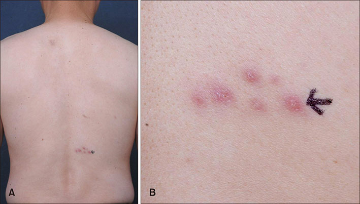
Fig. 1 (A) Multiple grouped erythematous papules mimicking herpes infection on the back of a 53-year-old man and (B) biopsy site (black arrow).
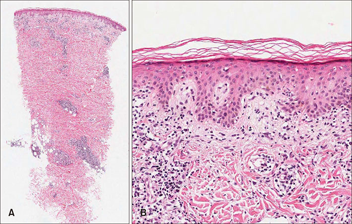
Fig. 2 (A) Lymphohistiocytic infiltration along dermoepidermal junction and around appendages, from superficial to deep dermis (H&E, ×40). (B) Hydropic changes in epidermal basal layer and mild mucin deposition in dermis (H&E, ×200).
Reference
-
1. Obermoser G, Sontheimer RD, Zelger B. Overview of common, rare and atypical manifestations of cutaneous lupus erythematosus and histopathological correlates. Lupus. 2010; 19:1050–1070.
Article2. Costner MI, Sontheimer RD, Provost TT. Lupus erythematosus. In : Sontheimer RD, Provost TT, editors. Cutaneous manifestations of rheumatic diseases. 2nd ed. Philadelphia: Lippincott Williams and Wilkins;2004. p. 15–64.3. Lee NY, Daniel AS, Dasher DA, Morrell DS. Cutaneous lupus after herpes zoster: isomorphic, isotopic, or both? Pediatr Dermatol. 2013; 30:e110–e113.
Article4. Longhi BS, Centeville M, Marini R, Appenzeller S. Koebner's phenomenon in systemic lupus erythematosus. Rheumatol Int. 2012; 32:1403–1405.
Article
