J Korean Ophthalmol Soc.
2018 Dec;59(12):1185-1189. 10.3341/jkos.2018.59.12.1185.
A Case of Isolated Traumatic Aniridia in a Pseudophakic Eye
- Affiliations
-
- 1Department of Ophthalmology, Daegu Fatima Hospital, Daegu, Korea. dj_oph_2540@hanmail.net
- KMID: 2428692
- DOI: http://doi.org/10.3341/jkos.2018.59.12.1185
Abstract
- PURPOSE
We report a rare case of isolated traumatic aniridia in a pseudophakic eye.
CASE SUMMARY
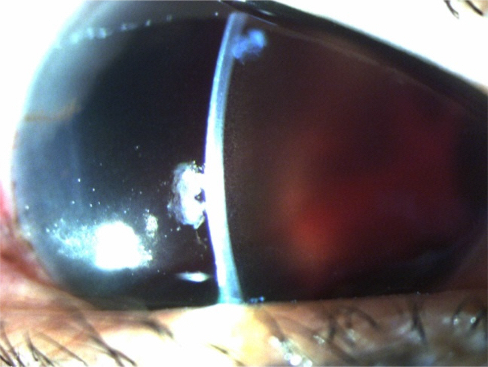
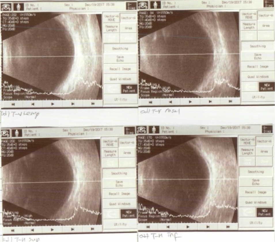
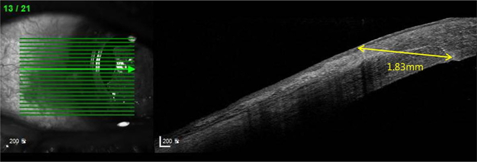
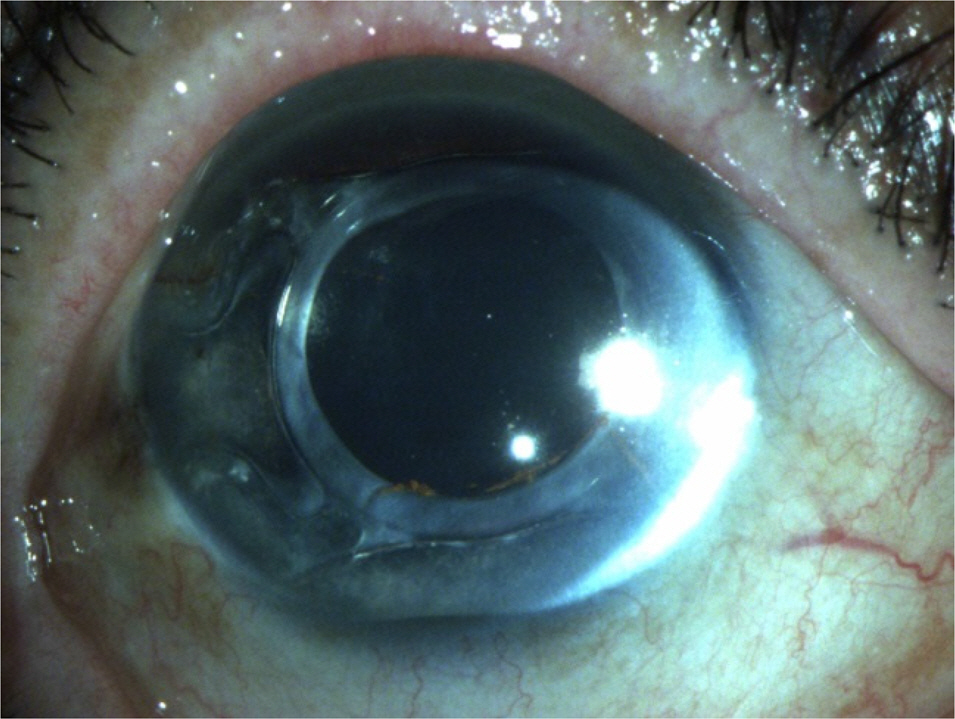
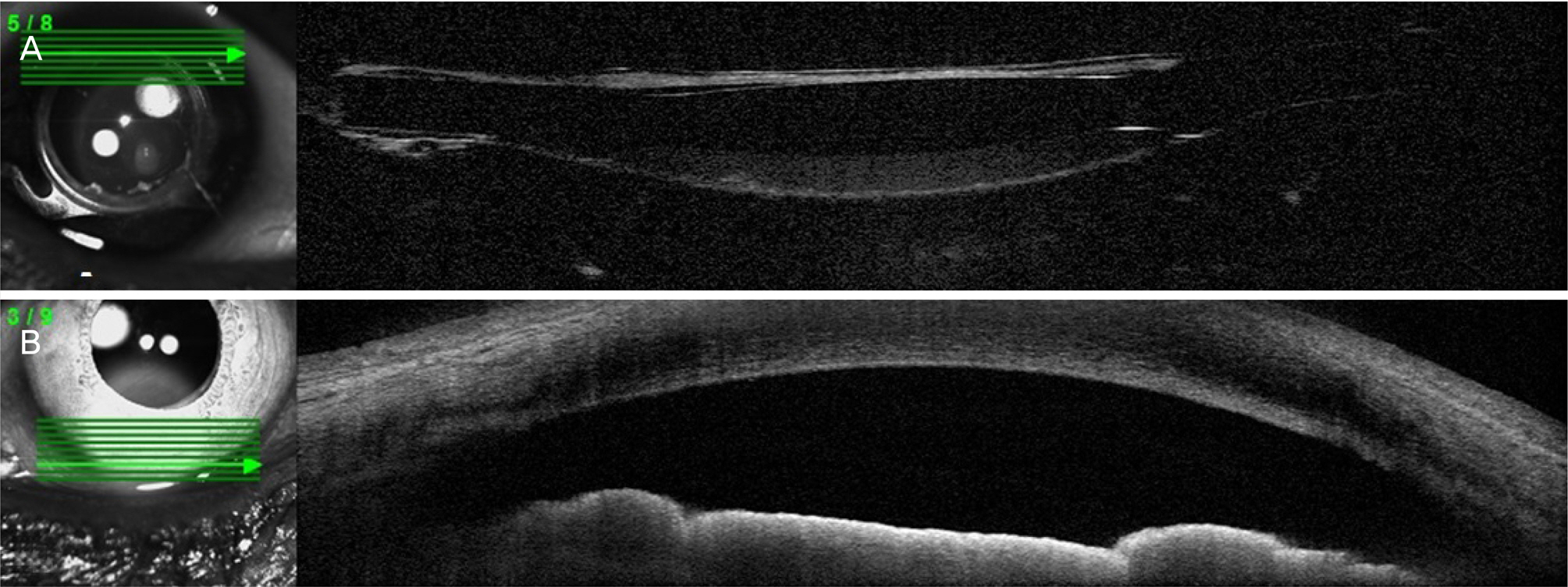
A 69-year-old female came to our emergency department complaining of right eye pain and visual disturbance after trauma due to fall on the stairs. Five years earlier she had undergone an uncomplicated right sutureless phacoemulsification cataract extraction through a 2.2 mm temporal clear corneal incision, followed by insertion of a folding intracapsular intraocular lens. Total iris expulsion occurred through the cataract incision without extension of the wound or disruption of the posterior capsule or intraocular lens.
CONCLUSIONS
We report a rare case of isolated traumatic aniridia in a pseudophakic eye, which has not been reported in the Republic of Korea.
Keyword
MeSH Terms
Figure
Reference
-
References
1. Kim HJ, Kwon JY. A clinical observation of perforating ocular injuries. J Korean Ophthalmol Soc. 1989; 30:123–30.2. Kwon JY, Cho HI. Perforating eye injuries in children. J Korean Ophthalmol Soc. 1984; 25:701–6.3. Romem M, Singer L. Traumatic aniridia. Br J Ophthalmol. 1973; 57:613–4.
Article4. Song KS, Yi GY. A case of traumatic complete aniridia with abdominal laceration. J Korean Ophthalmol Soc. 2000; 41:2013–7.5. Kass MA, Lahav M. Albert DM. Traumatic rupture of healed abdominal wounds. Am J Ophthalmol. 1976; 81:722–4.6. Magargal LE, Shakin E, Bolling JP, Robb-Doyle E. Traumatic abdominal of posterior chamber lenses: clinical and experimental correlations. J Cataract Refract Surg. 1986; 12:670–3.7. Johns KJ, Sheils P, Parrish CM, et al. Traumatic wound dehiscence in pseudophakia. Am J Ophthalmol. 1989; 108:535–9.
Article8. Assia EI, Blotnick CA, Powers TP, et al. Clinicopathologic study of ocular trauma in eyes with intraocular lenses. Am J Ophthalmol. 1994; 117:30–6.
Article9. Navon SE. Expulsive iridodialysis: an isolated injury after phacoemulsification. J Cataract Refract Surg. 1997; 23:805–7.
Article10. Lim JI, Nahl A, Johnston R, Jarus G. Traumatic total iridectomy due to iris extrusion through a self-sealing cataract incision. Arch Ophthalmol. 1999; 117:542–3.11. Ball J, Caesar R, Choudhuri D. Mystery of the vanishing iris. J Cataract Refract Surg. 2002; 28:180–1.
Article12. Prabhu A, Nayak H, Palimar P. Traumatic expulsive aniridia after phacoemulsification. Indian J Ophthalmol. 2007; 55:232–3.
Article13. Sullivan CA, Murray A, McDonnel P. The long-term results of nonexpulsive total iridodialysis: an isolated injury after phacoemu lsification. Eye (Lond). 2004; 18:534–6.14. Allan BD. Mechanism of iris prolapse: a qualitative analysis and implications for surgical technique. J Cataract Refract Surg. 1995; 21:182–6.
Article15. Muzaffar W, O'Duffy D. Traumatic aniridia in a pseudophakic eye. J Cataract Refract Surg. 2006; 32:361–2.
Article16. Ball JL, McLeod BK. Traumatic wound dehiscence following abdominal surgery: a thing of the past? Eye (Lond). 2001; 15(Pt 1):42–4.

