Bioabsorbable Screws Used in Hallux Valgus Treatment Using Proximal Chevron Osteotomy
- Affiliations
-
- 1Department of Orthopedics, Inje University Sanggye Paik Hospital, Seoul, Korea.
- 2Department of Orthopedic Surgery, Gwangju Veterans Hospital, Gwangju, Korea. mdaky@hanmail.net
- KMID: 2428661
- DOI: http://doi.org/10.14193/jkfas.2018.22.4.181
Abstract
- Hallux valgus is a deformity that causes pain in the first metatarsophalangeal joint. Surgical methods are quite diverse and a range of osteotomies are used at the proximal and distal part of the metatarsal bone and proximal phalange. Fixation methods, such as plate, screw, K-wire, and others have been used in various ways. The fixation device is often removed with various side effects due to the fixation devices. In the case of instruments that are absorbed in vivo, these procedures are not necessary to remove and there is an advantage of not performing the second operation. Three patients were treated, in which a proximal chevron osteotomy was used with a bioabsorbable screw (K-METâ„¢; U&I Corporation).
MeSH Terms
Figure
-

Figure 1. Bioabsorbable headless screw (K-METTM; U&I Corporation).
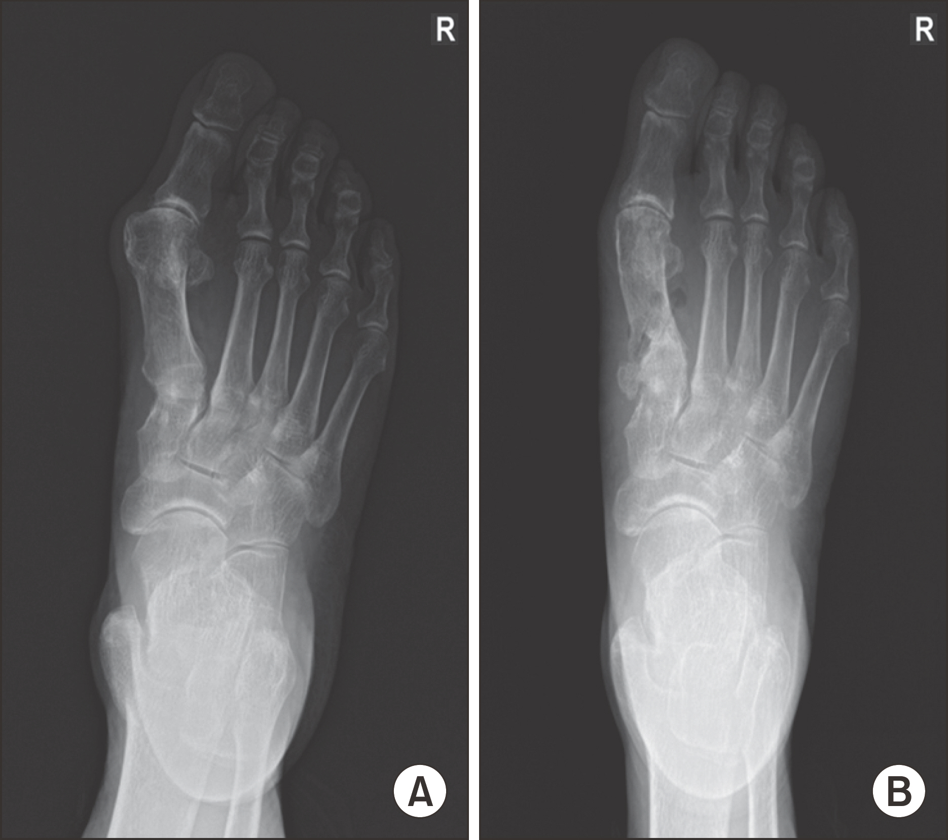
Figure 2. A 73-year-old male hallux valgus deformity patient shows hallux valgus angle 38。 and intermetatarsal angle 15。 in preoperation (A) and after 1 month hallux valgus angle 15。 and intermetatarsal angle 7。 in postoperation (B).

Figure 3. A 80-year-old female hallux valgus deformity patient shows hallux valgus angle 51。 and intermetatarsal angle 16。 in preoperation (A) and after 1 month hallux valgus angle 25。 and intermetatarsal angle 9。 in postoperation (B).
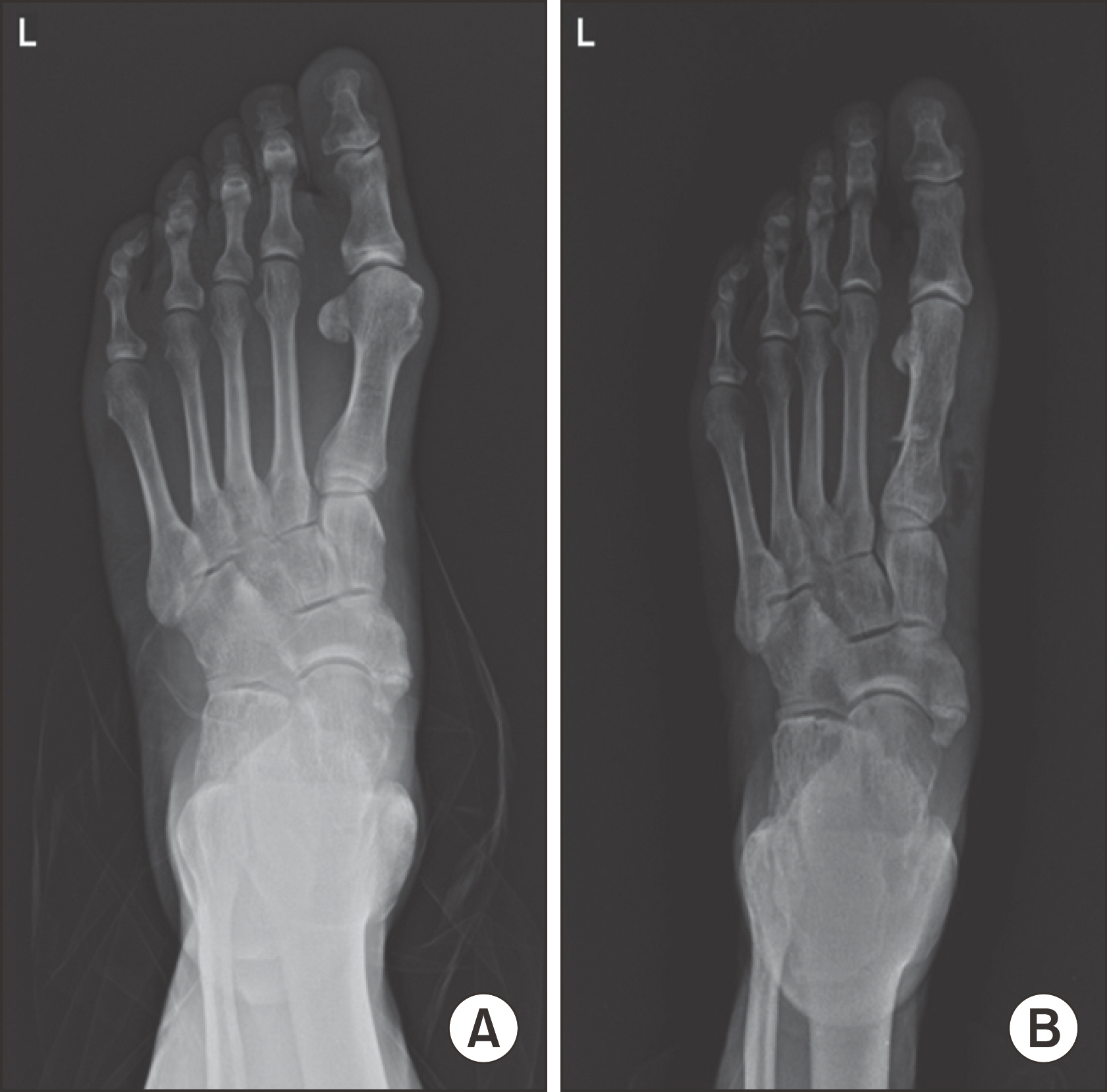
Figure 4. 76-year-old male hallux valgus deformity patient shows hallux valgus angle 36。 and intermetatarsal angle 14。 in preoperation (A) and after 1 month hallux valgus angle 11。 and intermetatarsal angle 6。 in postoperation (B).
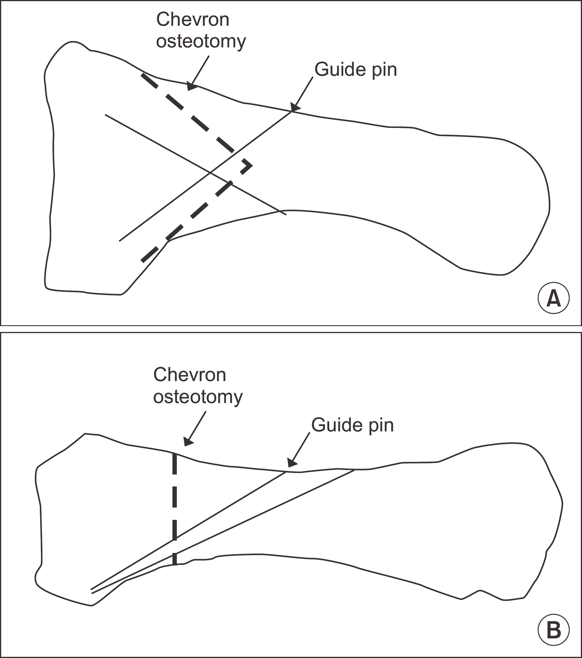
Figure 5. Guide pin insertion direction on 1st metatarsal bone. (A) Lateral view. (B) Anteroposterior view.
Reference
-
1.Kim JS., Cho HK., Young KW., Lee SY., Kim JS., Lee K. Effectiveness of headless bioabsorbable screws for fixation of the scarf osteotomy. Foot Ankle Int. 2016. 37:1189–96.
Article2.Park BJ., An KY., Choi YS. Arthroscopic assisted bioabsorbable screw fixation for radial head fractures: a report of two cases. J Korean Fract Soc. 2017. 30:35–9.
Article3.Easley ME., Kiebzak GM., Davis WH., Anderson RB. Prospective, randomized comparison of proximal crescentic and proximal chevron osteotomies for correction of hallux valgus deformity. Foot Ankle Int. 1996. 17:307–16.
Article4.Crosby LA., Bozarth GR. Fixation comparison for chevron osteotomies. Foot Ankle Int. 1998. 19:41–3.
Article5.Anderson RB., Davis WH. Internal fixation of the proximal chevron osteotomy. Foot Ankle Int. 1997. 18:371–2.
Article6.Bozkurt M., Tigaran C., Dalstra M., Jensen NC., Linde F. Stability of a cannulated screw versus a Kirschner wire for the proximal crescentic osteotomy of the first metatarsal: a biomechanical study. J Foot Ankle Surg. 2004. 43:138–43.
Article7.Lee JW., Han HS., Han KJ., Park J., Jeon H., Ok MR, et al. Long-term clinical study and multiscale analysis of in vivo biodegradation mechanism of Mg alloy. Proc Natl Acad Sci U S A. 2016. 113:716–21.
Article8.Kim TS., Kim HJ., Park YH., Lim HT. The differences of fixation method in proximal chevron osteotomy for Hallux Valgus: K-Wire, Cannulated screw, plate. J Korean Foot Ankle Soc. 2011. 15:62–7.9.Burns AE., Varin J. Poly-L-lactic and rod fixation results in foot surgery. J Foot Ankle Surg. 1998. 37:37–41.10.Kim YM., Cho BK., Kim DS., Choi ES., Shon HC., Park KJ, et al. The distal metatarsal dorsal-wedge osteotomy using bio-compression screw for advanced Hallux Rigidus. J Korean Foot Ankle Soc. 2012. 16:38–46.
- Full Text Links
-
- Actions
-
Cited
- CITED
-
- Close
- Share
-
- Similar articles
-
- Chevron Osteotomy as the Treatment of Moderate to Severe Hallux Valgus Deformity
- A Comparison of Operative Treatment of Hallux Valgus with a Proximal Metatarsal Osteotomy and with a Modified Chevron Osteotomy
- Treatment of Severe Hallux Valgus Deformity with Proximal Reverse Chevron Metatarsal Osteotomy and Akin Osteotomy
- Proximal Metatarsal Chevron Osteotomy Combined with Modified McBride Procedures for Hallux Valgus Patients.
- Comparison of Proximal Metatarsal Osteotomy andDistal Chevron Osteotomy for Correction of Hallux Valgus
