Non-osseous Coalition of the Third Metatarsal-Lateral Cuneiform Joint: A Case Report
- Affiliations
-
- 1Department of Orthopedic Surgery, Seoul Medical Center, Seoul, Korea. 1435man@hanmail.net
- KMID: 2428660
- DOI: http://doi.org/10.14193/jkfas.2018.22.4.177
Abstract
- This paper reports a rare case of the symptomatic third metatarsal (MT3) - lateral cuneiform (LC) in a 55-year-old male who presented with complaints of severe intermittent pain in his right foot. Plain radiographs and computed tomography scans revealed sclerosis and irregularity at this joint. The intraoperative findings demonstrated a fibrocartilaginous coalition. The pain had improved one year after removing the MT3-LC joint by en bloc and arthrodesis.
Figure
-
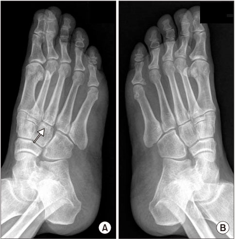
Figure 1 Medial oblique radiograph demonstrates the third metatarsal-lateral cuneiform joint irregularity (arrow) of the right foot (A) joint compared to left foot oblique view (B).
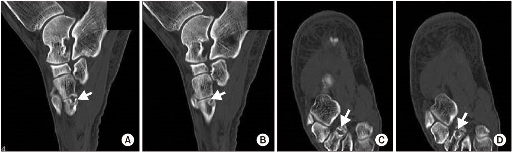
Figure 2 Computed tomography scans of the right foot demonstrates cortical irregularity (A, C) and subchondral cyst (B, D) of the third metatarsal-lateral cuneiform joint (arrows).
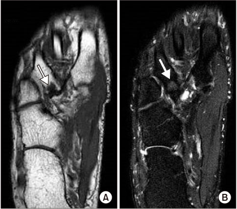
Figure 3 Magnetic resonance imaging scans of the right foot demonstrates low signal in T1-weighted image (A) (arrow) and intermediate signal in T2-weighted image (B) (arrow) of the third metatarsal-lateral cuneiform joint.
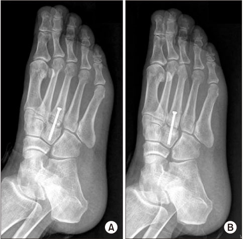
Figure 4 Postoperative radiographs of the right the third metatarsal-lateral cuneiform joint demonstrates non-union on first trial (A) and second trial (B).
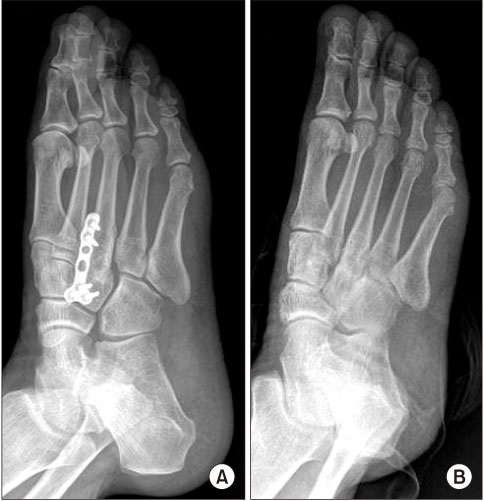
Figure 5 (A, B) Postoperative radiographs of the right the third metatarsal-lateral cuneiform joint demonstrates union on third trial.
Reference
-
1. Day FN 3rd, Naples JJ, White J. Metatarsocuneiform coalition. J Am Podiatr Med Assoc. 1994; 84:197–199.
Article2. Coughlin MJ, Mann RA, Saltzman CL. Surgery of the foot and ankle. Volume II. 8th ed. Philadelphia: Mosby;2007. p. 1073–1082.3. Stevens BW, Kolodziej P. Non-osseous tarsal coalition of the lateral cuneiform-third metatarsal joint. Foot Ankle Int. 2008; 29:867–870.
Article4. Regan MH, Case DT, Brundige JC. Articular surface defects in the third metatarsal and third cuneiform: nonosseous tarsal coalition. Am J Phys Anthropol. 1999; 109:53–65.
Article5. Conway JJ, Cowell HR. Tarsal coalition: clinical significance and roentgenographic demonstration. Radiology. 1969; 92:799–811.
Article6. Kumai T, Takakura Y, Akiyama K, Higashiyama I, Tamai S. Histopathological study of nonosseous tarsal coalition. Foot Ankle Int. 1998; 19:525–531.
Article7. Ouzounian TJ, Shereff MJ. In vitro determination of midfoot motion. Foot Ankle. 1989; 10:140–146.
Article8. Takakura Y, Nakata H. Isolated first cuneometatarsal coalition: a case report. Foot Ankle Int. 1999; 20:815–817.
Article9. Tanaka Y, Takakura Y, Sugimoto K, Kumai T. Non-osseous coalition of the medial cuneiform-first metatarsal joint: a case report. Foot Ankle Int. 2000; 21:1043–1046.
Article10. Comfort TK, Johnson LO. Resection for symptomatic talocalcaneal coalition. J Pediatr Orthop. 1998; 18:283–288.
Article
- Full Text Links
-
- Actions
-
Cited
- CITED
-
- Close
- Share
-
- Similar articles
-
- Bilateral Naviculo-Medial Cuneiform Coalition: One Case Report
- Subtalar Coalition: Usefulness of the C Sign on Lateral Radiographs of the Ankle
- Naviculo-Medial Cuneiform Coalition: Radiological Features
- Unilateral Talonavicular Coalition: A Case Report
- Operative Treatment of Symptomatic Naviculocuneiform Coalition in Children: 2 Cases Report
