Int J Thyroidol.
2018 May;11(1):56-59. 10.11106/ijt.2018.11.1.56.
A Case of Zenker's Diverticulum Mimicking a Right Side Thyroid Nodule
- Affiliations
-
- 1Air Defense & Control Command, ROKAF, Seoul, Korea.
- 2Division of Endocrinology, Department of Internal Medicine, Chung-Ang University College of Medicine, Seoul, Korea. hyahnmd@cau.ac.kr
- KMID: 2428072
- DOI: http://doi.org/10.11106/ijt.2018.11.1.56
Abstract
- Zenker's diverticulum, a pulsion diverticulum of the hypopharynx, is a rare lesion that commonly occurs in left side of hypopharynx. The incidence of esophageal diverticula is much lower than that of focal lesions or nodules of thyroid. In an ultrasonography, the outpouching just like a focal thyroid lesion, may present as an oval or circular structure. The food remnants or gas bubbles present in the diverticulum may mimic microcalcifications presented in papillary thyroid carcinoma. We reported a case of right side Zenker's diverticulum mimicking a thyroid cancer in thyroid ultrasonography.
Keyword
MeSH Terms
Figure
-
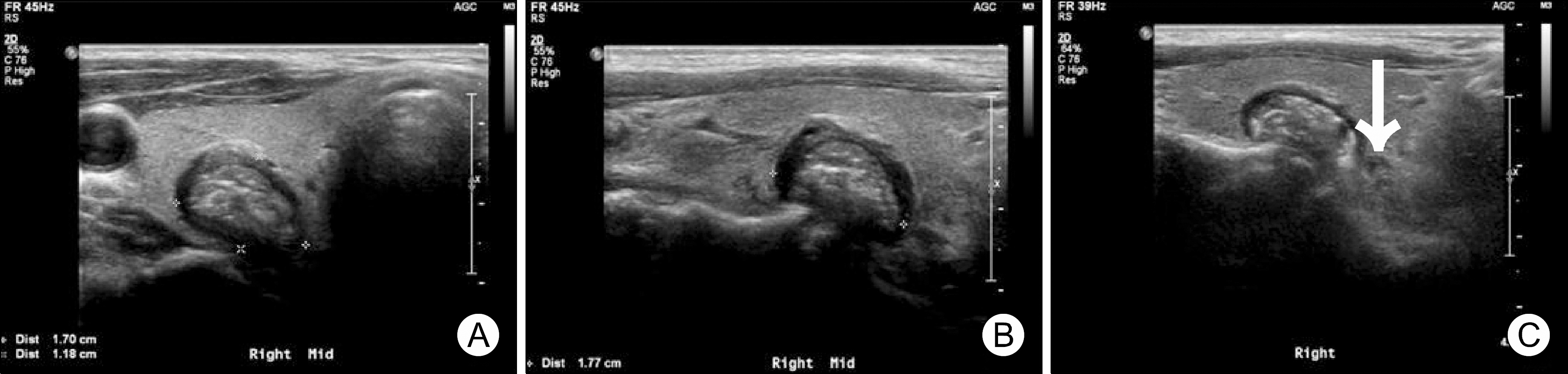
Fig. 1. The axial (A) and longitudinal (B) US scan of right thyroid lobe shows 17×12×18 mm sized isoechoic nodule with micro-and macrocalcification. The arrow shows communication of nodule and esophagus (C).
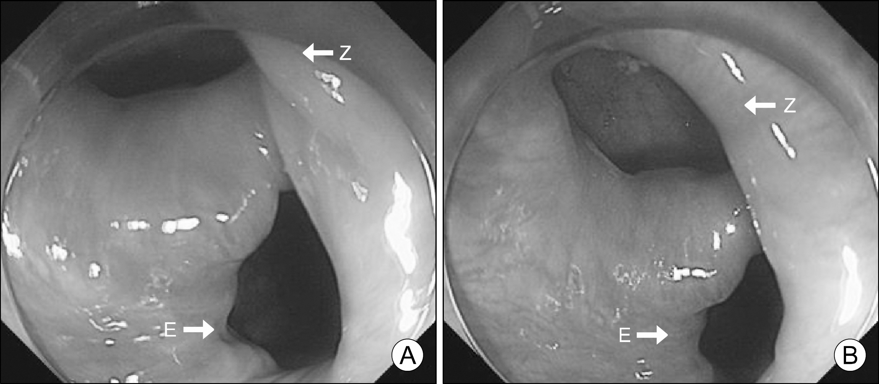
Fig. 2. (A, B) This esophago-gastroscopy shows an outpouching lesion of upper esophagus. E: esophagus, Z: Zenker’ s diverticulum
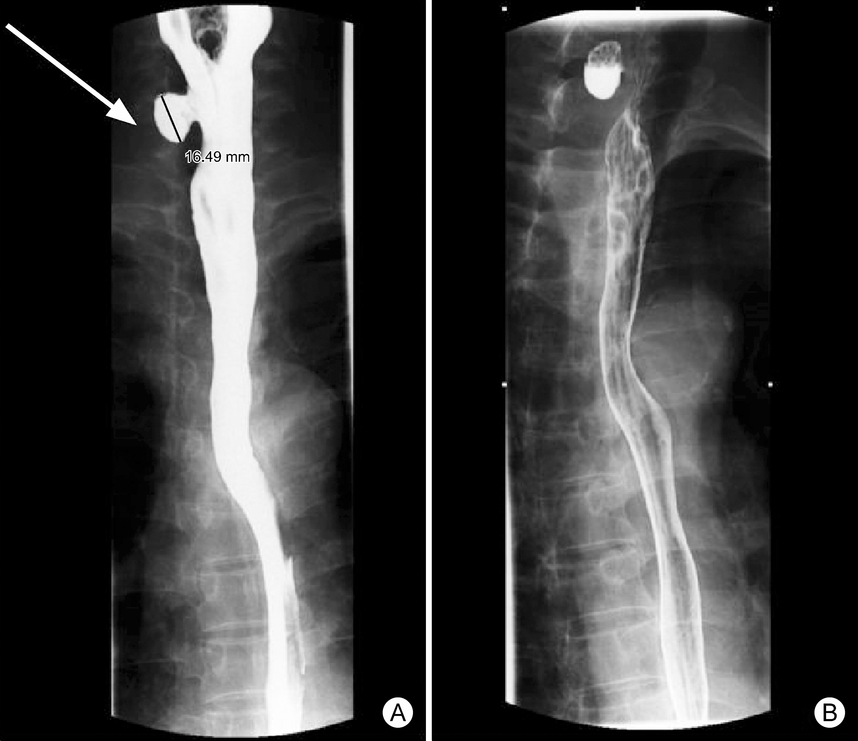
Fig. 3. The esophagography confirms a barium filled sac projecting from the upper esophagus, posterior aspects.(A) Barium filling view, (B) mucosal relief view. Arrow: Zenker's diverticulum.
Reference
-
1). Law R, Katzka DA, Baron TH. Zenker's diverticulum. Clin Gastroenterol Hepatol. 2014; 12(11):1773–82. quiz e111-2.
Article2). Kim HK, Lee JI, Jang HW, Bae SY, Lee JH, Kim YS, et al. Characteristics of Killian-Jamieson diverticula mimicking a thyroid nodule. Head Neck. 2012; 34(4):599–603.
Article3). Kim SJ, Kim CH. The genetic studies of obsessive-compulsive disorder and its future directions. Yonsei Med J. 2006; 47(4):443–54.
Article4). Haugen BR, Alexander EK, Bible KC, Doherty GM, Mandel SJ, Nikiforov YE, et al. 2015 American Thyroid Association Management Guidelines for adult patients with thyroid nodules and differentiated thyroid cancer: The American Thyroid Association Guidelines Task Force on thyroid nodules and differentiated thyroid cancer. Thyroid. 2016; 26(1):1–133.
Article5). Yoon HD, Shon HS. Killian-Jamieson diverticulum mimicking a thyroid nodule. Korean J Med. 2005; 68(4):467–8.6). DeFriend DE, Dubbins PA. Sonographic demonstration of a pharyngoesophageal diverticulum. J Clin Ultrasound. 2000; 28(9):485–7.
Article7). Komatsu M, Komatsu T, Inove K. Ultrasonography of Zenker's diverticulum: special reference to differential diagnosis from thyroid nodules. Eur J Ultrasound. 2000; 11(2):123–5.
Article8). Stafford ND, Moore-Gillon V, McKelvie P. Handedness and the side on which pharyngeal pouches occur. Br Med J (Clin Res Ed). 1984; 288(6420):815–6.
Article9). Cao L, Ge J, Zhao D, Lei S. Killian-Jamieson diverticulum mimicking a calcified thyroid nodule on ultrasonography: a case report and literature review. Oncol Lett. 2016; 12(4):2742–5.
Article10). Mimatsu K, Oida T, Kano H, Kawasaki A, Fukino N, Kida K, et al. Killian-jamieson diverticula presenting synchronously with thyroid adenoma. Case Rep Gastroenterol. 2013; 7(1):188–94.
Article11). Pang JC, Chong S, Na HI, Kim YS, Park SJ, Kwon GY. Killian-Jamieson diverticulum mimicking a suspicious thyroid nodule: sonographic diagnosis. J Clin Ultrasound. 2009; 37(9):528–30.
Article12). Kwak JY, Kim EK. Sonographic findings of Zenker diverticula. J Ultrasound Med. 2006; 25(5):639–42.
Article
- Full Text Links
-
- Actions
-
Cited
- CITED
-
- Close
- Share
-
- Similar articles
-
- A Case of Zenker’s Diverticulum Mimicking a Thyroid Nodule
- A Case of Killian-Jamieson Diverticulum Mimicking a Thyroid Nodule
- A Case of Zenker's Diverticulum Mimicking the Thyroid Tumor Associated with Vocal Cord Palsy
- Incidentally Found Pharyngoesophageal Diverticulum on Ultrasonography
- A Case of Papillary Thyroid Carcinoma Accompanied by Zenker’s Diverticulum: Considerations for Surgery
