Intramuscular Epidermal Cyst of the Buttock: A Case Report
- Affiliations
-
- 1Department of Radiology, Konkuk University School of Medicine, Seoul, Korea. kimnr001@gmail.com
- 2Department of Radiology, Gangwon National University Hospital, Chuncheon, Korea.
- KMID: 2427385
- DOI: http://doi.org/10.3348/jksr.2018.79.6.354
Abstract
- Epidermal cysts are common benign subcutaneous lesions that occur in or on the skin. It is not very difficult to diagnose subcutaneous epidermal cysts using ultrasound imaging because they exhibit typical sonographic features. However, the differential diagnosis can be confused when epidermal cysts are found in unusual sites. The authors report a case involving a 4-year-old girl who presented with an intramuscular epidermal cyst in the gluteus maximus muscle. Magnetic resonance imaging revealed characteristic internal features of the epidermal cyst, despite being in an uncommon site, and was very useful in the preoperative diagnosis.
MeSH Terms
Figure
-
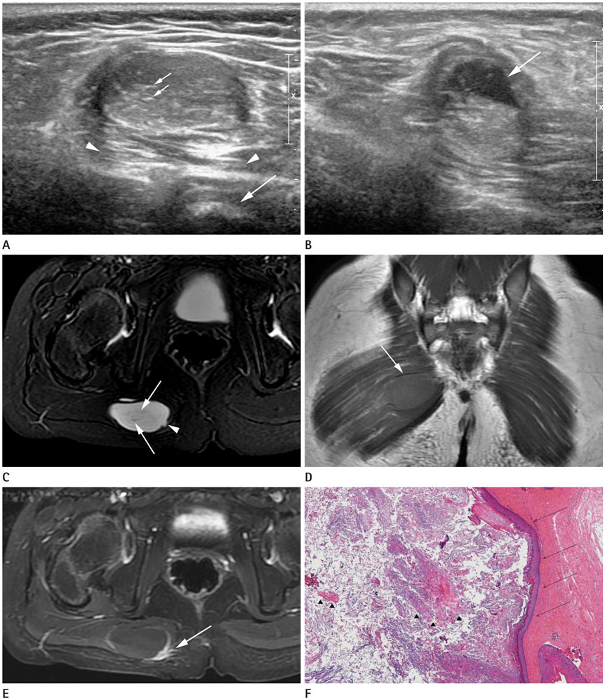
Fig. 1 Intramuscular Epidermal Cyst of the buttock in a 4-year-old girl presented with a palpable mass in the right buttock. A. Ultrasound imaging revealing an ovoid hyperechoic mass with internal hyperechoic strands (short arrows) and posterior acoustic enhancement (arrowheads) in the gluteus maximus muscle on transverse scan. Acoustic shadowing by the ischial tuberosity (long arrow) is noted. B. Ten months later, a follow-up ultrasound reveals an increase in the size of the mass, with extension to the subcutaneous tissue. The anechoic cystic change is indicated at a focal bulging portion to the subcutaneous layer (arrow). C. Axial T2-weighted magnetic resonance image revealing a bright, hyperintense cystic mass in the right gluteus maximus muscle. The cyst includes internal hypointense debris (arrows) and the appearance of focal budding (arrowhead). D. Coronal T1-weighted magnetic resonance image revealing a slightly high-intensity signal of the mass at the inferomedial aspect of the buttock (arrow). E. Gadolinium-enhanced fat-suppressed T1-weighted image revealing peripheral rim enhancement and focal adjacent soft tissue enhancement (arrow). F. Hematoxylin and eosin stained microscopic section revealing several layers of slightly atrophic squamous epithelial-lining cyst (black arrows) filled with horny materials (arrowheads). Pathological findings were consistent with epidermal cyst (× 200).
Reference
-
1. Yang DM, Yoon MH, Kim HS, Oh YH, Ha SY, Oh JH, et al. Presacral epidermoid cyst: imaging findings with histopathologic correlation. Abdom Imaging. 2001; 26:79–82.
Article2. Lever WH, Lever GS. Tumors and cysts of epidermis. In : Elder D, editor. Histopathology of the skin. 8th ed. Philadelphia, JB: Lippincott-Raven;1997. p. 685–746.3. Elder D, Elenitsas R, Jaworsky C, Johnson JB. Lever's histopathology of the skin. 8th ed. Philadelphia: Lippincott-Raven;1997. p. 695–721.4. Patel K, Bhuiya T, Chen S, Kenan S, Kahn L. Epidermal inclusion cyst of phalanx: a case report and review of the literature. Skeletal Radiol. 2006; 35:861–863.
Article5. Garg M, Kataria SP, Sethi D, Mathur SK. Epidermoid cyst of spleen mimicking splenic lymphangioma. Adv Biomed Res. 2013; 2:49.
Article6. Chatterjee PK, Chandra AB, Dastidar N. Epidermal cyst in sternomastoid muscle simulating a malignant growth. Indian J Otolaryngol Head Neck Surg. 1976; 28:86–87.7. Ozawa T, Harada T, Ishii M. Giant epidermal cyst extending from sole to dorsum of the foot by penetrating the interosseous muscles. J Dermatol. 2008; 35:25–28.
Article8. Low SF, Sridharan R, Ngiu CS. Giant epidermal cyst with intramuscular extension: a rare occurrence. BMJ Case Rep. 2015; 2015:bcr2013202534.
Article9. Kim HK, Kim SM, Lee SH, Racadio JM, Shin MJ. Subcutaneous epidermal inclusion cysts: ultrasound (US) and MR imaging findings. Skeletal Radiol. 2011; 40:1415–1419.
Article10. Hong SH, Chung HW, Choi JY, Koh YH, Choi JA, Kang HS. MRI findings of subcutaneous epidermal cysts: emphasis on the presence of rupture. AJR Am J Roentgenol. 2006; 186:961–966.
Article
