Anatomical Variants of Lister's Tubercle: A New Morphological Classification Based on Magnetic Resonance Imaging
- Affiliations
-
- 1Department of Radiology, Changi General Hospital, Singapore 529889, Singapore. le_roy_chong@cgh.com.sg
- KMID: 2427205
- DOI: http://doi.org/10.3348/kjr.2017.18.6.957
Abstract
OBJECTIVE
Lister's tubercle is used as a standard anatomical landmark in hand surgery and arthroscopy procedures. In this study, we aimed to evaluate and propose a classification for anatomical variants of Lister's tubercle.
MATERIALS AND METHODS
Between September 2011 and July 2014, 360 MRI examinations for wrists performed using 1.5T scanners in a single institution were retrospectively evaluated. The prevalence of anatomical variants of Lister's tubercle based on the heights and morphology of its radial and ulnar peaks was assessed. These were classified into three distinct types: radial peak larger than ulnar peak (Type 1), similar radial and ulnar peaks (Type 2) and ulnar peak larger than radial peak (Type 3). Each type was further divided into 2 subtypes (A and B) based on the morphology of the peaks.
RESULTS
The proportions of Type 1, Type 2, and Type 3 variants in the study population were 69.2, 21.4, and 9.5%, respectively. For the subtypes, the Type 1A variant was the most common (41.4%) and conformed to the classical appearance of Lister's tubercle; whereas, Type 3A and 3B variants were rare configurations (6.4% and 3.1%, respectively) wherein the extensor pollicis longus tendon coursed along the radial aspect of Lister's tubercle.
CONCLUSION
Anatomical variations of Lister's tubercle have potential clinical implications for certain pathological conditions and pre-procedural planning. The proposed classification system facilitates a better understanding of these anatomical variations and easier identification of at-risk and rare variants.
MeSH Terms
Figure
-
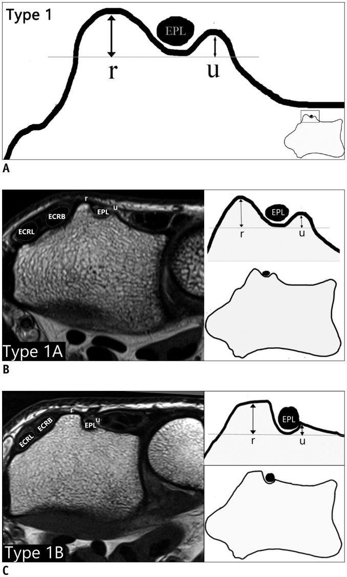
Fig. 1 Type 1 variant on diagrams and axial MR images.A. Schematic of Type 1 variant. B. Type 1A variant (classical configuration of Lister's tubercle). C. Type 1B variant wherein radial (r) peak is dominant with box-like configuration and ulnar peak (u) is small. ECRB = extensor carpi radialis brevis, ECRL = extensor carpi radialis longus, EPL = extensor pollicis longus

Fig. 2 Type 2 variant on diagrams and axial MR images.A. Schematic of Type 2 variant. B. Type 2A variant wherein both radial (r) and ulnar (u) peaks are discrete and equal in height. C. Type 2B variant wherein both radial (r) and ulnar (u) peaks are equal in height but rudimentary and difficult to visualize.
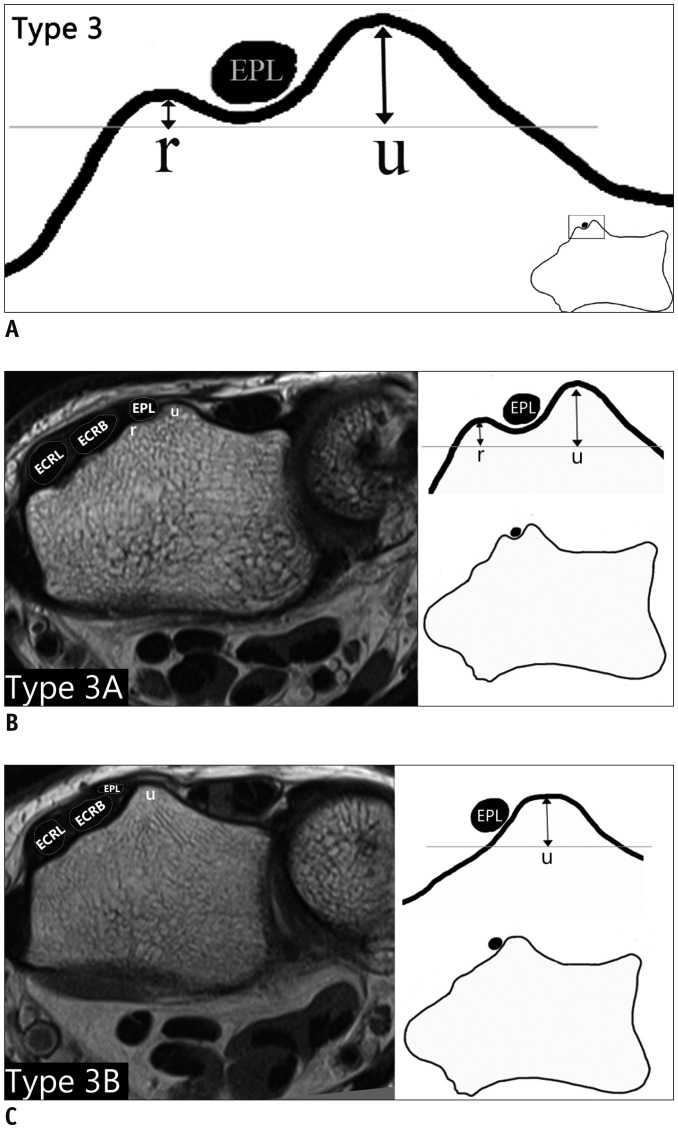
Fig. 3 Type 3 variant on diagrams and axial MR images.A. Schematic of Type 3 variant. B. Type 3A variant wherein the ulnar (u) peak is dominant and taller than radial (r) peak. C. Type 3B variant wherein ulnar (u) peak is dominant with absent radial (r) peak. In these variants, EPL tendon appears to cross radial to palpated Lister's tubercle.
Cited by 1 articles
-
Age of Data in Contemporary Research Articles Published in Representative General Radiology Journals
Ji Hun Kang, Dong Hwan Kim, Seong Ho Park, Jung Hwan Baek
Korean J Radiol. 2018;19(6):1172-1178. doi: 10.3348/kjr.2018.19.6.1172.
Reference
-
1. Saffar P, Fakhoury B. [Secondary repair of the extensor pollicis longus]. Ann Chir Main. 1987; 6:225–229. PMID: 3426331.2. Hazani R, Engineer NJ, Cooney D, Wilhelmi BJ. Anatomic landmarks for the first dorsal compartment. Eplasty. 2008; 8:e53. PMID: 19092992.3. Smith J, Rizzo M, Finnoff JT, Sayeed YA, Michaud J, Martinoli C. Sonographic appearance of the posterior interosseous nerve at the wrist. J Ultrasound Med. 2011; 30:1233–1239. PMID: 21876094.
Article4. Ikiz ZA, Uçerler H. Anatomic characteristics and clinical importance of the superficial branch of the radial nerve. Surg Radiol Anat. 2004; 26:453–458. PMID: 15365770.
Article5. Smith DK. Dorsal carpal ligaments of the wrist: normal appearance on multiplanar reconstructions of threedimensional Fourier transform MR imaging. AJR Am J Roentgenol. 1993; 161:119–125. PMID: 8517289.
Article6. Lohman M, Vasenius J, Nieminen O. Ultrasound guidance for puncture and injection in the radiocarpal joint. Acta Radiol. 2007; 48:744–747. PMID: 17729005.
Article7. Slutsky DJ. Wrist arthroscopy. In : Wolfe SW, Hotchkiss RN, Pederson WC, Kozin SH, editors. Green's operative hand surgery. 6th ed. Philadelphia: Elsevier;2011. p. 709–741.8. Berger RA. A method of defining palpable landmarks for the ligament-splitting dorsal wrist capsulotomy. J Hand Surg Am. 2007; 32:1291–1295. PMID: 17923317.
Article9. Barbieri CH, Mazzer N. Triradiate skin incision for dorsal approach to the wrist. J Hand Surg Br. 1996; 21:21–23. PMID: 8676023.
Article10. Samarakoon LB, Lakmal KC, Thillainathan S, Bataduwaarachchi VR, Anthony DJ, Jayasekara RW. Anatomical relations of the superficial sensory branches of the radial nerve: a cadaveric study with clinical implications. Patient Saf Surg. 2011; 5:28. PMID: 22054296.
Article11. Galvis EJ, Kumar KK, Özyurekoglu T. Radioscapholunate arthrodesis using low-profile dorsal pi plate. Tech Hand Up Extrem Surg. 2013; 17:80–83. PMID: 23689853.
Article12. Espen D. [Fixation of fractures of the distal radius using the “nail-plate”]. Oper Orthop Traumatol. 2009; 21:459–471. PMID: 20058124.13. Hochwald NL, Levine R, Tornetta P 3rd. The risks of Kirschner wire placement in the distal radius: a comparison of techniques. J Hand Surg Am. 1997; 22:580–584. PMID: 9260610.
Article14. Pichler W, Windisch G, Schaffler G, Rienmüller R, Grechenig W. Computer tomography aided 3D analysis of the distal dorsal radius surface and the effects on volar plate osteosynthesis. J Hand Surg Eur Vol. 2009; 34:598–602. PMID: 19959446.
Article15. Ağr I, Aytekin MN, Küçükdurmaz F, Gökhan S, Cavus¸ UY. Anatomical localization of Lister’s tubercle and its clinical and surgical importance. Open Orthop J. 2014; 8:74–77. PMID: 24843388.16. Kaplan EB. Anatomical variations of the forearm and hand. In : Tubiana R, editor. The hand. Philadelphia: W. B. Saunders;1981. p. 361–374.17. Valentin P. Extrinsic muscles of the hand and wrist: an introduction. In : Tubiana R, editor. The hand. Philadelphia: WB Saunders;1981. p. 243.18. Clement H, Pichler W, Nelson D, Hausleitner L, Tesch NP, Grechenig W. Morphometric analysis of Lister’s tubercle and its consequences on volar plate fixation of distal radius fractures. J Hand Surg Am. 2008; 33:1716–1719. PMID: 19084168.
Article19. Benson EC, DeCarvalho A, Mikola EA, Veitch JM, Moneim MS. Two potential causes of EPL rupture after distal radius volar plate fixation. Clin Orthop Relat Res. 2006; 451:218–222. PMID: 16770281.
Article20. Maschke SD, Evans PJ, Schub D, Drake R, Lawton JN. Radiographic evaluation of dorsal screw penetration after volar fixed-angle plating of the distal radius: a cadaveric study. Hand (N Y). 2007; 2:144–150. PMID: 18780076.
Article21. Ozer K, Toker S. Dorsal tangential view of the wrist to detect screw penetration to the dorsal cortex of the distal radius after volar fixed-angle plating. Hand (N Y). 2011; 6:190–193. PMID: 22654703.
Article22. Lamas C, Llusà M, Méndez A, Proubasta I, Carrera A, Forcada P. Intraosseous vascularity of the distal radius: anatomy and clinical implications in distal radius fractures. Hand (N Y). 2009; 4:418–423. PMID: 19475457.
Article23. Shima H, Ohno K, Michi K, Egawa K, Takiguchi R. An anatomical study on the forearm vascular system. J Craniomaxillofac Surg. 1996; 24:293–299. PMID: 8938512.
Article24. Lanzetta M, Howard M, Conolly WB. Post-traumatic triggering of extensor pollicis longus at the dorsal radial tubercle. J Hand Surg Br. 1995; 20:398–401. PMID: 7561421.
Article25. Stahl S, Wolff TW. Delayed rupture of the extensor pollicis longus tendon after nonunion of a fracture of the dorsal radial tubercle. J Hand Surg Am. 1988; 13:338–341. PMID: 3379265.
Article26. Ferreres A, Llusá M, García-Elías M, Lluch A. A possible mechanism of direct injury to the EPL tendon at Lister's tubercle during falls with the wrist fully extended. J Hand Surg Eur Vol. 2008; 33:149–151. PMID: 18443053.27. Rowland P, Phelan N, Gardiner S, Linton KN, Galvin R. The effectiveness of corticosteroid injection for de Quervain’s stenosing tenosynovitis (DQST): A Systematic Review and meta-analysis. Open Orthop J. 2015; 9:437–444. PMID: 26587059.
Article28. Nishijo K, Kotani H, Miki T, Senzoku F, Ueo T. Unusual course of the extensor pollicis longus tendon associated with tenosynovitis, presenting as de Quervain disease--a case report. Acta Orthop Scand. 2000; 71:426–428. PMID: 11028897.
Article
- Full Text Links
-
- Actions
-
Cited
- CITED
-
- Close
- Share
-
- Similar articles
-
- Radiologic Analysis of Distal Radius Fracture Accompanying Spontaneous Extensor Pollicis Longus Rupture
- Ultrafast Magnetic Resonance Imaging: Echo Planar Imaging and Spiral Scan Imaging
- Functional Magnetic Resonance Imaging of the Brain: Principle and Practical Application
- Computed Tomography-Based Morphologic Analysis of Osteoarthritis of the Distal Radioulnar Joint Associated with Extensor Tendon Ruptures
- Radiologic Evaluation and Differential Diagnosis of Gallbladder Cancer
